Difference between revisions of "Basal cell carcinoma"
Jump to navigation
Jump to search
(→References: +EL) |
|||
| Line 75: | Line 75: | ||
DDx: | DDx: | ||
*Benign hair follicle - lacks necrosis, core has lower cellular density, surrounding sheath composed of loose connective tissue. | |||
*[[Trichoepithelioma]] - no artefactual cleft,<ref name=Ref_PBoD8_1180-1>{{Ref PBoD8|1180-1}}</ref> and typically no [[solar elastosis]]. | *[[Trichoepithelioma]] - no artefactual cleft,<ref name=Ref_PBoD8_1180-1>{{Ref PBoD8|1180-1}}</ref> and typically no [[solar elastosis]]. | ||
*[[Adenoid cystic carcinoma]] - no myxoid stroma, no peripheral palisading. | *[[Adenoid cystic carcinoma]] - no myxoid stroma, no peripheral palisading. | ||
Revision as of 15:11, 12 September 2013
| Basal cell carcinoma | |
|---|---|
| Diagnosis in short | |
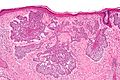
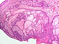
 Basal cell carcinoma. H&E stain. | |
|
| |
| LM | "basaloid cells", nests with peripheral palisading of cells, artefactual clefting, myxoid stroma |
| Subtypes | superficial pattern, nodular pattern, morpheaform (sclerosing) pattern, infiltrative pattern, fibroepitheliomatous pattern, infundibulocystic pattern, adenoidal pattern |
| LM DDx | trichoepithelioma, adenoid cystic carcinoma, eccrine poroma, reticulated seborrheic keratosis (for BCC, fibroepitheliomatous pattern), basaloid squamous cell carcinoma, basosquamous carcinoma |
| Gross | pearly nodule with telangiectasias |
| Site | skin |
|
| |
| Syndromes | Bazex syndrome, nevoid basal cell carcinoma syndrome, xeroderma pigmentosum |
|
| |
| Prevalence | very common |
| Prognosis | good |
| Clin. DDx | solar elastosis with ectatic blood vessels |
| Basal cell carcinoma | |
|---|---|
| External resources | |
| EHVSC | 10187 (BCC with dermal scar from bx) |
Basal cell carcinoma, abbreviated BCC, is an extremely common form of skin cancer.
General
- Very common.
- Sun exposed skin.
- Hair bearing area; tumour derived from hair follicle - a more appropriate name might be trichoblastic carcinoma.[1]
- Very rarely metastasizes:
- Dermatopathologists might see a couple in their career.
- There are only ~ 300 literature reports of metastatic BCC.[2]
Clinical
- Telangiectasias.
- Raised pearly nodule.
As part of a syndrome
- Nevoid basal cell carcinoma syndrome (NBCCS), AKA Gorlin syndrome.
- Bazex syndrome (X-linked).[3]
- Xeroderma pigmentosum.
Microscopic
- Basaloid cells - similar in appearance to basal cells:
- Moderate blue/grey cytoplasm.
- Dark ovoid/ellipsoid nucleus with uniform chromatin.
- Palisading of cells at the edge of the cell nests.
- Artefactual separation of cells (forming the nests) from the underlying stroma - key feature.
- Surrounded by blue (myxoid) stroma - key feature.
May be present:[5]
- Dystrophic calcification - possibly more aggressive behaviour.[6]
- Amyloid.
- Inflammation.
Notes:
- Palisading = the long axes of the cells are alined and the axes are perpendicular to the interface between the (basaloid cell) nests and stroma.
- Key elements in a list: Artefactual clefting (of nests), Basaloid cells, Peripheral palisading, Myxoid stroma.
- Memory device PAM: palisading, artefactual clefts, myxoid stroma.
DDx:
- Benign hair follicle - lacks necrosis, core has lower cellular density, surrounding sheath composed of loose connective tissue.
- Trichoepithelioma - no artefactual cleft,[4] and typically no solar elastosis.
- Adenoid cystic carcinoma - no myxoid stroma, no peripheral palisading.
- Eccrine poroma - on palms & soles, BCC rarely found there.[7]
- Reticulated seborrheic keratosis - for BCC, fibroepitheliomatous pattern.
- Basaloid squamous cell carcinoma - AKA squamous cell carcinoma, basaloid variant.
- Basosquamous carcinoma - squamous cell carcinoma with basal cell carcinoma (a collision tumour).
- Solar elastosis with ectatic blood vessels.
Images
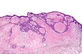
BCC - intermed. mag. (WC/Nephron)
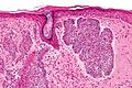
BCC - high mag. (WC/Nephron)
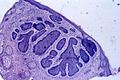
BCC - poor quality. (WC)
Fibroepithelioma of Pinkus. (WC/Nephron)
Fibroepithelioma of Pinkus. (WC)


www:
- BCC (ucsf.edu).[8]
- BCC with fibroepitheliomatous pattern / fibroepithelioma of Pinkus (surgicalpathologyatlas.com).
- BCC with fibroepitheliomatous pattern (dermatlas.med.jhmi.edu).
- BCC in vitiligo (nih.gov).[9]
Basal cell carcinoma subtypes/unique features
- Many patterns exist.
- Recurrence higher in morpheaform (sclerosing), infiltrative, micronodular, and superficial patterns.[10]
- DG says the prognosis is similar for all BCC subtypes, except for sclerosing pattern and infiltrative pattern.[11]
The subtypes:[12]
| Pattern | Key histologic feature | Other histologic features | Other |
|---|---|---|---|
| Superficial pattern | connected to epidermis | ||
| Nodular pattern | nodules | partial detachment from epidermis | subgroup micronodular = nests equal size ~ 0.2 mm dia., >=25% of lesion |
| Morpheaform (sclerosing) pattern | stroma sclerosis | often seen with infiltrative pattern, DDx: desmoplastic trichoepithelioma[13] | |
| Infiltrative pattern | small irregular cell aggregates | often also sclerosing or morpheaform | |
| Fibroepitheliomatous pattern | cords and columns of basaloid cells | fibrous stroma | name of pattern comes from fibroepithelioma of Pinkus; DDx: reticulated seborrheic keratosis |
| Infundibulocystic pattern | small keratocysts (keratin cysts) | usu. small, often in cords | usu. indolent |
| Adenoidal pattern | cribriform / pseudoglandular arch. | myxoid stroma, peripheral palisading | DDx: adenoid cystic carcinoma |
Unique features/differentiation:[12]
| Differentiation / unique cell | Key histologic feature | Other histologic features | Other |
|---|---|---|---|
| Pigmented cells | any pattern can have pigmentation | pigment may be in malignant cell | DDx: collision lesion with melanocytic lesion |
| Squamous differentiation (metatypical BCC) | pink cytoplasm, keratinization | assoc. with ulceration/tumour recurrence | |
| Eccrine differentiation | focal duct formation | very rare, DDx: BCC engulfing sweat ducts | |
| Clear cells (Clear cell BCC) | clear cytoplasm | due to glycogen |
IHC
- CK5/6 +ve.
- Useful to assess margins... if very close.
- CD10 +ve.
- Actin +ve.
Squamous cell carcinoma versus basal cell carcinoma:
- BerEP4 +ve.
- SCC usually negative.[14]
- EMA -ve.
- SCC usually positive.[15]
- SMA +ve.[16]
- SCC usually negative.
Sign-out
SKIN LESION, SHAVE BIOPSY WITH ELECTRODESICCATION AND CURETTAGE (EDC):
- BASAL CELL CARCINOMA, MARGIN STATUS ASSESSED CLINICALLY DURING EDC.
- EXTENSIVE SOLAR ELASTOSIS.
SKIN LESION, RIGHT EAR, EXCISION:
- BASAL CELL CARCINOMA.
- MARGINS NEGATIVE FOR BASAL CELL CARCINOMA.
- EXTENSIVE SOLAR ELASTOSIS.
SKIN LESION, RIGHT TEMPLE, RE-EXCISION:
- BASAL CELL CARCINOMA, NODULAR, MARGINS NEGATIVE.
- DERMAL SCAR.
- EXTENSIVE SOLAR ELASTOSIS.
Micro
The sections show hair-bearing skin with nests of basaloid cells in the dermis. The basaloid nests have peripheral palisading of the nuclei, have numerous mitoses, and are surrounded by a myxoid stroma. The nests are well demarcated from the stroma and show focal clefting from the stroma. The margins are negative for basal cell carcinoma.
See also
References
- ↑ Busam, Klaus J. (2009). Dermatopathology: A Volume in the Foundations in Diagnostic Pathology Series (1st ed.). Saunders. pp. 389. ISBN 978-0443066542.
- ↑ Ting, PT.; Kasper, R.; Arlette, JP. (Jan 2005). "Metastatic basal cell carcinoma: report of two cases and literature review.". J Cutan Med Surg 9 (1): 10-5. doi:10.1007/s10227-005-0027-1. PMID 16208438.
- ↑ URL: http://emedicine.medscape.com/article/1101146-diagnosis. Accessed on: 6 May 2010.
- ↑ 4.0 4.1 Kumar, Vinay; Abbas, Abul K.; Fausto, Nelson; Aster, Jon (2009). Robbins and Cotran pathologic basis of disease (8th ed.). Elsevier Saunders. pp. 1180-1. ISBN 978-1416031215.
- ↑ 5.0 5.1 Busam, Klaus J. (2009). Dermatopathology: A Volume in the Foundations in Diagnostic Pathology Series (1st ed.). Saunders. pp. 390. ISBN 978-0443066542.
- ↑ Slodkowska, EA.; Cribier, B.; Peltre, B.; Jones, DM.; Carlson, JA. (Aug 2010). "Calcifications associated with basal cell carcinoma: prevalence, characteristics, and correlations.". Am J Dermatopathol 32 (6): 557-64. doi:10.1097/DAD.0b013e3181ca65e2. PMID 20489568.
- ↑ Tadrous, Paul.J. Diagnostic Criteria Handbook in Histopathology: A Surgical Pathology Vade Mecum (1st ed.). Wiley. pp. 284. ISBN 978-0470519035.
- ↑ URL: http://missinglink.ucsf.edu/lm/DermatologyGlossary/basal_cell_carcinoma.html. Accessed on: 4 September 2011.
- ↑ Rustemeyer, J.; Günther, L.; Deichert, L. (Sep 2011). "A rare association: basal cell carcinoma in a vitiliginous macula.". Oral Maxillofac Surg 15 (3): 175-7. doi:10.1007/s10006-010-0240-y. PMID 20623309.
- ↑ Basal cell carcinoma. eMedicine. Prognosis section. URL: http://emedicine.medscape.com/article/276624-overview. Accessed on: 17 September 2011.
- ↑ Ghazarian, Danny; 14 September 2011.
- ↑ 12.0 12.1 Busam, Klaus J. (2009). Dermatopathology: A Volume in the Foundations in Diagnostic Pathology Series (1st ed.). Saunders. pp. 392-5. ISBN 978-0443066542.
- ↑ Kirzhner, M.; Jakobiec, FA.; Borodic, G.. "Desmoplastic trichoepithelioma: report of a unique periocular case.". Ophthal Plast Reconstr Surg 28 (5): e121-3. doi:10.1097/IOP.0b013e318245535a. PMID 22366669.
- ↑ Yu, L.; Galan, A.; McNiff, JM. (Oct 2009). "Caveats in BerEP4 staining to differentiate basal and squamous cell carcinoma.". J Cutan Pathol 36 (10): 1074-176. doi:10.1111/j.1600-0560.2008.01223.x. PMID 19187107.
- ↑ Beer, TW.; Shepherd, P.; Theaker, JM. (Sep 2000). "Ber EP4 and epithelial membrane antigen aid distinction of basal cell, squamous cell and basosquamous carcinomas of the skin.". Histopathology 37 (3): 218-23. PMID 10971697.
- ↑ URL: http://www.ihcworld.com/_newsletter/2004/2004-12_basal_cell_vs_squamous_v1.pdf. Accessed on: 19 December 2012.