Difference between revisions of "Barrett's esophagus"
Jump to navigation
Jump to search
(redirect) |
(split-out) |
||
| Line 1: | Line 1: | ||
:''Intestinal metaplasia of the esophagus'' redirects here. | |||
'''Barrett esophagus''', abbreviated '''BE''', is a relatively common pathology of the esophagus that is associated with an increased risk of [[esophageal adenocarcinoma]]. | |||
==General== | |||
*Diagnosis is made by '''clinicans ''not'' pathologists'''. | |||
**A common histologic correlate is metaplastic transformation of stratified squamous epithelium to simple columnar epithelium with goblet cells. | |||
***There is disagreement whether goblet cells are required for the diagnosis.<ref name=pmid19623166>{{Cite journal | last1 = Riddell | first1 = RH. | last2 = Odze | first2 = RD. | title = Definition of Barrett's esophagus: time for a rethink--is intestinal metaplasia dead? | journal = Am J Gastroenterol | volume = 104 | issue = 10 | pages = 2588-94 | month = Oct | year = 2009 | doi = 10.1038/ajg.2009.390 | PMID = 19623166 }}</ref> | |||
****One large study suggests that goblets cells are only absent due to undersampling.<ref name=pmid21959311>{{Cite journal | last1 = Chandrasoma | first1 = P. | last2 = Wijetunge | first2 = S. | last3 = DeMeester | first3 = S. | last4 = Ma | first4 = Y. | last5 = Hagen | first5 = J. | last6 = Zamis | first6 = L. | last7 = DeMeester | first7 = T. | title = Columnar-lined esophagus without intestinal metaplasia has no proven risk of adenocarcinoma. | journal = Am J Surg Pathol | volume = 36 | issue = 1 | pages = 1-7 | month = Jan | year = 2012 | doi = 10.1097/PAS.0b013e31822a5a2c | PMID = 21959311 }}</ref> | |||
*Associated with (chronic) [[gastroesophageal reflux disease]]. | |||
Significance of Barrett's esophagus: | |||
*Increased risk of adenocarcinoma of the esophagus. | |||
**Need on-going surveillance, i.e. long term follow-up/repeat esophagogastroduodenoscopy. | |||
==Gross== | |||
*Red/light brown esophageal mucosa. | |||
**Normal mucosa = light pink. | |||
<gallery> | |||
Image:Barretts_esophagus.jpg | Endoscopic image of BE. (WC) | |||
</gallery> | |||
==Microscopic== | |||
Features: | |||
*Columnar epithelium with: | |||
**Goblet cells - '''key feature'''. | |||
**+/-Moderate chronic inflammation +/- acute inflammation -- common.<ref name=pmid10566710>{{Cite journal | last1 = Voutilainen | first1 = M. | last2 = Färkkilä | first2 = M. | last3 = Mecklin | first3 = JP. | last4 = Juhola | first4 = M. | last5 = Sipponen | first5 = P. | title = Chronic inflammation at the gastroesophageal junction (carditis) appears to be a specific finding related to Helicobacter pylori infection and gastroesophageal reflux disease. The Central Finland Endoscopy Study Group. | journal = Am J Gastroenterol | volume = 94 | issue = 11 | pages = 3175-80 | month = Nov | year = 1999 | doi = 10.1111/j.1572-0241.1999.01513.x | PMID = 10566710 }}</ref> | |||
**+/-Mild nuclear hyperchromasia. | |||
*+/-Squamous epithelium with changes of [[gastroesophageal reflux disease|gastroesophageal reflux]]. | |||
DDx: | |||
*[[Chronic gastritis]]. | |||
*[[Helicobacter gastritis]]. | |||
*[[Low-grade columnar dysplasia of the esophagus]]. | |||
===Images=== | |||
<gallery> | |||
Image:Barretts_alcian_blue.jpg | Barrett's type mucosa. [[Alcian blue stain]]. (WC) | |||
Image:Barrett's_mucosa,_PAS-Alcian_blue_stain.jpg | Barrett's type mucosa. Alcian blue stain. (WC/AFIP) | |||
Image:Barrett's_mucosa,_higher_magnification,_Alcian_blue_stain_.jpg | Barrett's type mucosa. Alcian blue stain. (WC/AFIP) | |||
</gallery> | |||
==Stains== | |||
*Alcian blue (pH 2.5)<ref name=pmid10517897>{{Cite journal | last1 = Voutilainen | first1 = M. | last2 = Färkkilä | first2 = M. | last3 = Juhola | first3 = M. | last4 = Mecklin | first4 = JP. | last5 = Sipponen | first5 = P. | title = Complete and incomplete intestinal metaplasia at the oesophagogastric junction: prevalences and associations with endoscopic erosive oesophagitis and gastritis. | journal = Gut | volume = 45 | issue = 5 | pages = 644-8 | month = Nov | year = 1999 | doi = | PMID = 10517897 |URL = http://gut.bmj.com/content/45/5/644.full }}</ref> - goblet cells +ve. | |||
==Sign-out== | |||
<pre> | |||
ESOPHAGUS, DISTAL, BIOPSY: | |||
- COLUMNAR EPITHELIUM WITH INTESTINAL METAPLASIA AND MILD ACUTE INFLAMMATION, SEE COMMENT. | |||
- REACTIVE SQUAMOUS EPITHELIUM. | |||
- NEGATIVE FOR DYSPLASIA AND NEGATIVE FOR MALIGNANCY. | |||
COMMENT: | |||
The findings are consistent with Barrett's esophagus in the appropriate endoscopic setting. | |||
</pre> | |||
<pre> | |||
ESOPHAGUS, DISTAL, BIOPSY: | |||
- COLUMNAR EPITHELIUM WITH INTESTINAL METAPLASIA AND MODERATE CHRONIC INFLAMMATION, SEE COMMENT. | |||
- REACTIVE SQUAMOUS EPITHELIUM. | |||
- NEGATIVE FOR DYSPLASIA AND MALIGNANCY. | |||
COMMENT: | |||
The findings are consistent with Barrett's esophagus in the appropriate endoscopic setting. | |||
</pre> | |||
<pre> | |||
ESOPHAGUS, DISTAL, BIOPSY: | |||
- COLUMNAR EPITHELIUM WITH EXTENSIVE INTESTINAL METAPLASIA, ACUTE AND CHRONIC INFLAMMATION; | |||
- SEE COMMENT. | |||
- REACTIVE SQUAMOUS EPITHELIUM. | |||
- NEGATIVE FOR DYSPLASIA AND MALIGNANCY. | |||
COMMENT: | |||
The columnar epithelium with intestinal metplasia is seen located deep to the squamous | |||
epithelium. | |||
The findings are consistent with Barrett's esophagus in the appropriate endoscopic setting. | |||
</pre> | |||
==See also== | |||
*[[Esophagus]] | |||
*[[GERD]]. | |||
==References== | |||
{{Reflist|2}} | |||
[[Category:Esophagus]] | |||
[[Category:Diagnosis]] | [[Category:Diagnosis]] | ||
Revision as of 03:15, 24 October 2013
- Intestinal metaplasia of the esophagus redirects here.
Barrett esophagus, abbreviated BE, is a relatively common pathology of the esophagus that is associated with an increased risk of esophageal adenocarcinoma.
General
- Diagnosis is made by clinicans not pathologists.
- A common histologic correlate is metaplastic transformation of stratified squamous epithelium to simple columnar epithelium with goblet cells.
- Associated with (chronic) gastroesophageal reflux disease.
Significance of Barrett's esophagus:
- Increased risk of adenocarcinoma of the esophagus.
- Need on-going surveillance, i.e. long term follow-up/repeat esophagogastroduodenoscopy.
Gross
- Red/light brown esophageal mucosa.
- Normal mucosa = light pink.
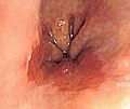
Endoscopic image of BE. (WC)

Microscopic
Features:
- Columnar epithelium with:
- Goblet cells - key feature.
- +/-Moderate chronic inflammation +/- acute inflammation -- common.[3]
- +/-Mild nuclear hyperchromasia.
- +/-Squamous epithelium with changes of gastroesophageal reflux.
DDx:
Images
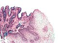
Barrett's type mucosa. Alcian blue stain. (WC)
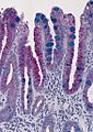
Barrett's type mucosa. Alcian blue stain. (WC/AFIP)
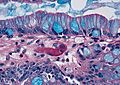
Barrett's type mucosa. Alcian blue stain. (WC/AFIP)


Stains
- Alcian blue (pH 2.5)[4] - goblet cells +ve.
Sign-out
ESOPHAGUS, DISTAL, BIOPSY:
- COLUMNAR EPITHELIUM WITH INTESTINAL METAPLASIA AND MILD ACUTE INFLAMMATION, SEE COMMENT.
- REACTIVE SQUAMOUS EPITHELIUM.
- NEGATIVE FOR DYSPLASIA AND NEGATIVE FOR MALIGNANCY.

COMMENT:
The findings are consistent with Barrett's esophagus in the appropriate endoscopic setting.
ESOPHAGUS, DISTAL, BIOPSY:
- COLUMNAR EPITHELIUM WITH INTESTINAL METAPLASIA AND MODERATE CHRONIC INFLAMMATION, SEE COMMENT.
- REACTIVE SQUAMOUS EPITHELIUM.
- NEGATIVE FOR DYSPLASIA AND MALIGNANCY.

COMMENT:
The findings are consistent with Barrett's esophagus in the appropriate endoscopic setting.
ESOPHAGUS, DISTAL, BIOPSY:
- COLUMNAR EPITHELIUM WITH EXTENSIVE INTESTINAL METAPLASIA, ACUTE AND CHRONIC INFLAMMATION;
- SEE COMMENT.
- REACTIVE SQUAMOUS EPITHELIUM.
- NEGATIVE FOR DYSPLASIA AND MALIGNANCY.

COMMENT:
The columnar epithelium with intestinal metplasia is seen located deep to the squamous
epithelium.

The findings are consistent with Barrett's esophagus in the appropriate endoscopic setting.
See also
References
- ↑ Riddell, RH.; Odze, RD. (Oct 2009). "Definition of Barrett's esophagus: time for a rethink--is intestinal metaplasia dead?". Am J Gastroenterol 104 (10): 2588-94. doi:10.1038/ajg.2009.390. PMID 19623166.
- ↑ Chandrasoma, P.; Wijetunge, S.; DeMeester, S.; Ma, Y.; Hagen, J.; Zamis, L.; DeMeester, T. (Jan 2012). "Columnar-lined esophagus without intestinal metaplasia has no proven risk of adenocarcinoma.". Am J Surg Pathol 36 (1): 1-7. doi:10.1097/PAS.0b013e31822a5a2c. PMID 21959311.
- ↑ Voutilainen, M.; Färkkilä, M.; Mecklin, JP.; Juhola, M.; Sipponen, P. (Nov 1999). "Chronic inflammation at the gastroesophageal junction (carditis) appears to be a specific finding related to Helicobacter pylori infection and gastroesophageal reflux disease. The Central Finland Endoscopy Study Group.". Am J Gastroenterol 94 (11): 3175-80. doi:10.1111/j.1572-0241.1999.01513.x. PMID 10566710.
- ↑ Voutilainen, M.; Färkkilä, M.; Juhola, M.; Mecklin, JP.; Sipponen, P. (Nov 1999). "Complete and incomplete intestinal metaplasia at the oesophagogastric junction: prevalences and associations with endoscopic erosive oesophagitis and gastritis.". Gut 45 (5): 644-8. PMID 10517897.