Difference between revisions of "Ameloblastoma"
Jump to navigation
Jump to search
(split out) |
(+infobox) |
||
| Line 1: | Line 1: | ||
{{ Infobox diagnosis | |||
| Name = {{PAGENAME}} | |||
| Image = Ameloblastoma - high mag.jpg | |||
| Width = | |||
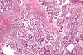
| Caption = Ameloblastoma. [[H&E stain]]. | |||
| Synonyms = | |||
| Micro = stellate reticulum (star-shaped cells), tall columnar cells that have palisaded nuclei with reverse polarization, subnuclear vacuolization, +/-giant cells, +/-subepithelial hyalinization (eosinophilic acellular amorphous material) | |||
| Subtypes = solid/multicystic, unicystic | |||
| LMDDx = [[adenomatoid odontogenic tumour]], [[ameloblastic fibroma]] | |||
| Stains = | |||
| IHC = | |||
| EM = | |||
| Molecular = | |||
| IF = | |||
| Gross = | |||
| Grossing = | |||
| Site = usu. mandible - see ''[[odontogenic tumours and cysts]]'' | |||
| Assdx = | |||
| Syndromes = | |||
| Clinicalhx = | |||
| Signs = | |||
| Symptoms = | |||
| Prevalence = uncommon | |||
| Bloodwork = | |||
| Rads = | |||
| Endoscopy = | |||
| Prognosis = | |||
| Other = | |||
| ClinDDx = [[keratocytic odontogenic tumour]], odontogenic cysts | |||
| Tx = | |||
}} | |||
'''Ameloblastoma''' is an [[odontogenic cyst]]. | '''Ameloblastoma''' is an [[odontogenic cyst]]. | ||
Revision as of 15:15, 23 February 2014
| Ameloblastoma | |
|---|---|
| Diagnosis in short | |
 Ameloblastoma. H&E stain. | |
|
| |
| LM | stellate reticulum (star-shaped cells), tall columnar cells that have palisaded nuclei with reverse polarization, subnuclear vacuolization, +/-giant cells, +/-subepithelial hyalinization (eosinophilic acellular amorphous material) |
| Subtypes | solid/multicystic, unicystic |
| LM DDx | adenomatoid odontogenic tumour, ameloblastic fibroma |
| Site | usu. mandible - see odontogenic tumours and cysts |
|
| |
| Prevalence | uncommon |
| Clin. DDx | keratocytic odontogenic tumour, odontogenic cysts |
Ameloblastoma is an odontogenic cyst.
General
- Osteous lesion.
- Usually mandible.[1]
- In a review of 3677 cases, the mandible-to-maxilla ratio was 5 to 1.[2]
- May arise from an odontogenic cyst,[3] e.g. dentigerous cyst.[4]
Classification
Location:
- Intra-osseous.
- Locally aggressive.
- Peripheral.
- Benign.
Subclassification of intra-osseous type
Histology:
- Solid/multicystic.
- More commonly reoccur.
- Unicystic.
- Unlikely to reoccur.
- Classically found in younger individuals.
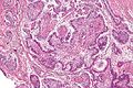
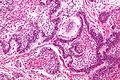
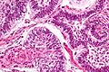
Microscopic
Features:[5]
- Stellate reticulum - star-shaped cells, found in a developing tooth.
- Tall columnar cells.
- Palisaded nuclei with reverse polarization.
- Reverse polarization of nuclei = nuclei distant from the basement membrane/nuclei at pole opposite of basement membrane.
- Palisaded nuclei = picket fence appearance; columnar-shaped nuclei with long axis perpendicular to the basement membrane -- key feature.
- Subnuclear vacuolization.
- Palisaded nuclei with reverse polarization.
- +/-Giant cells.
- +/-Subepithelial hyalinization (eosinophilic acellular amorphous material).
- Seen deep to the basement membrane.
- Variable morphology (see below - morphology).
DDx (nuclear palisading):
Images
www:
Ameloblastoma - low mag. (WC)
Ameloblastoma - intermed. mag. (WC)
Ameloblastoma - high mag. (WC)
Ameloblastoma - very high mag. (WC)


Morphology
- Not prognostic.
Morphologic variants:
- Follicular ameloblastoma (classic appearance).
- Plexiform ameloblastoma (does not have prominent palisading).
- Acanthomatous ameloblastoma.
- Desmoplastic ameloblastoma.
- Basaloid ameloblastoma.
See also
References
- ↑ URL: http://www.waent.org/archives/2010/Vol3-2/20100618-ameloblastoma/jaw-tumor.htm. Accessed on: 30 November 2011.
- ↑ Reichart, PA.; Philipsen, HP.; Sonner, S. (Mar 1995). "Ameloblastoma: biological profile of 3677 cases.". Eur J Cancer B Oral Oncol 31B (2): 86-99. PMID 7633291.
- ↑ Eversole, LR. (Nov 1999). "Malignant epithelial odontogenic tumors.". Semin Diagn Pathol 16 (4): 317-24. PMID 10587275.
- ↑ Moosvi, Z.; Tayaar, SA.; Kumar, GS. (Apr 2011). "Neoplastic potential of odontogenic cysts.". Contemp Clin Dent 2 (2): 106-9. doi:10.4103/0976-237X.83073. PMC 3180832. PMID 21957386. https://www.ncbi.nlm.nih.gov/pmc/articles/PMC3180832/.
- ↑ URL: http://www.pathconsultddx.com/pathCon/diagnosis?pii=S1559-8675%2806%2970616-7. Accessed on: March 9, 2010.