Multifocal micronodular pneumocyte hyperplasia associated with tuberous sclerosis
Jump to navigation
Jump to search
Multifocal micronodular pneumocyte hyperplasia associated with tuberous sclerosis, also multifocal micronodular pneumocyte hyperplasia in tuberous sclerosis, is the presence of a rare relatively distinctive hamartomatous lesion of the lung in multiple foci in a person with tuberous sclerosis.[1]
| Multifocal micronodular pneumocyte hyperplasia associated with tuberous sclerosis | |
|---|---|
| Diagnosis in short | |
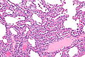
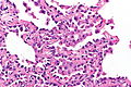
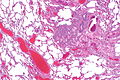
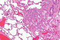
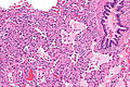
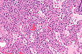
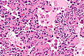
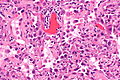
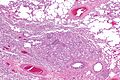
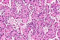
 Micronodule of pneumocyte hyperplasia in multifocal micronodular pneumocyte hyperplasia associated with tuberous sclerosis. H&E stain. | |
|
| |
| LM | enlarged alveolar lining cells with the hobnail morphology and round or oval nuclei, macrophages within the air spaces |
| LM DDx | atypical adenomatous hyperplasia of the lung |
| IHC | cytokeratin +ve, surfactant apoproteins (A & B) +ve, HMB-45 -ve |
| Molecular | mutations in TSC1 or TSC2 |
| Site | lung |
|
| |
| Associated Dx | lymphangioleiomyomatosis - also assoc. with tuberous sclerosis |
| Syndromes | tuberous sclerosis |
|
| |
| Prevalence | rare |
| Radiology | ground-glass nodules, +/-emphysematous changes |
| Prognosis | benign |
| Clin. DDx | multifocal AAH |
General
- Rare.
- May mimic multifocal atypical adenomatous hyperplasia on radiology.[2]
Clinical:
- May have recurrent pneumothorax.[3]
Gross
Features:[2]
- Multiple small lung nodules.
- Random distribution. ‡
- Up to 5 mm in size.
Radiology:
- May have an emphysema-like picture due to the obstruction of lymphatics and alveolar ducts from mass effect.[3]
- Nodules have ground-glass appearance on CT.[2]
Notes:
- ‡ One paper says peripheral location and upper lobe predominant.[1]
Microscopic
Features:
- Macrophages within the air spaces.
- Enlarged alveolar lining cells with:
- Hobnail morphology - free (luminal) surface area > attached/basal surface area.
- Round or oval nuclei.
DDx:
- Atypical adenomatous hyperplasia of the lung - usu. does not have macrophages within the air spaces.
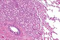
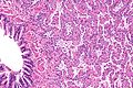
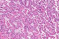
Images
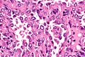
Set 1
MMPH - intermed. mag. (WC)
MMPH - high mag. (WC)

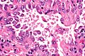
Set 2
MMPH - very low mag. (WC)
MMPH - low mag. (WC)
MMPH - intermed. mag. (WC)
MMPH - intermed. mag. (WC)
MMPH - high mag. (WC)
MMPH - high mag. (WC)


Set 3
MMPH - very low mag. (WC)
MMPH - low mag. (WC)
MMPH - intermed. mag. (WC)
MMPH - intermed. mag. (WC)
MMPH - high mag. (WC)
MMPH - very high mag. (WC)
MMPH - very high mag. (WC)


IHC
Features:[2]
- Cytokeratin +ve.
- Surfactant apoprotein A +ve.
- Surfactant apoprotein B +ve.
Others:[2]
- HMB-45 -ve.
- SMA (alpha) -ve.
- p53 -ve.
See also
References
- ↑ 1.0 1.1 Nagar, AM.; Teh, HS.; Khoo, RN.; Morani, AC.; Vrishni, K.; Raghuram, J. (Feb 2008). "Multifocal pneumocyte hyperplasia in tuberous sclerosis.". Thorax 63 (2): 186. doi:10.1136/thx.2006.076604. PMID 18234663.
- ↑ 2.0 2.1 2.2 2.3 2.4 Kobashi, Y.; Sugiu, T.; Mouri, K.; Irei, T.; Nakata, M.; Oka, M. (Jun 2008). "Multifocal micronodular pneumocyte hyperplasia associated with tuberous sclerosis: differentiation from multiple atypical adenomatous hyperplasia.". Jpn J Clin Oncol 38 (6): 451-4. doi:10.1093/jjco/hyn042. PMID 18535095.
- ↑ 3.0 3.1 Popper, HH.; Juettner-Smolle, FM.; Pongratz, MG. (Apr 1991). "Micronodular hyperplasia of type II pneumocytes--a new lung lesion associated with tuberous sclerosis.". Histopathology 18 (4): 347-54. PMID 2071093.