Coronary artery bypass grafts
Jump to navigation
Jump to search
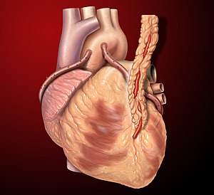
Coronary artery bypass grafts are a treatment for atherosclerotic heart disease (ASHD). They are created in a procedure called coronary artery bypass surgery, also coronary artery bypass grafting, abbreviated CABG. CABGs are encountered by pathologists at autopsy.
The other treatments for ASHD are medical management, and percutaneous coronary intervention (PCI).
Indications
Indications for CABG:[1]
- Left main coronary artery disease.
- Triple vessel disease.
- Disease involving the left anterior descending (coronary artery), left circumflex coronary artery and right coronary artery.
Others:
- Disease that cannot be treated with percutaneous coronary intervention (PCI).
Gross
.
Types of grafts
- Arterial - usually internal thoracic artery.
- Venous - saphenous vein.
Notes:
- Arterial grafts, generally, are much longer lived ~90% patency at 10 years verus ~57% for saphenous vein grafts.[2]
Common configurations
Arterial:
- Left internal thoracic artery (LITA) - almost always to the distal LAD.
Venous:
- Aorta to posterior interventricular branch of RCA.
- Typically pass around the right side of the heart.
- Aorta to left circumflex coronary artery.
- Typically pass around the left side of the heart.
- Aorta to first or second diagonal branch of the LAD.
Identification
- Know the common configurations.
- Get the OR note - important.
- Clinical notes are notoriously unreliable.
- Trace the grafts from proximal to distal or distal to proximal.
- Look for the sutures... where there is a suture there is often a graft.
- The aorta usu. has one extra suture for the (arterial) cannulation site -- where the bypass machine returns the blood to the body.
- The right atrium usu. has a suture for the (venous) cannulation site -- where the bypass machine receives the blood from the body.
All grafts should be identified:
- Proximal anastomosis - if applicable.
- Usu. non-existent in ITA grafts.
- Generally, found several centimetres above the coronary ostia.
- Distal anastomosis.
- Classic site of failure, esp. in arterial grafts.
See also
References
- ↑ Serruys, PW.; Morice, MC.; Kappetein, AP.; Colombo, A.; Holmes, DR.; Mack, MJ.; Ståhle, E.; Feldman, TE. et al. (Mar 2009). "Percutaneous coronary intervention versus coronary-artery bypass grafting for severe coronary artery disease.". N Engl J Med 360 (10): 961-72. doi:10.1056/NEJMoa0804626. PMID 19228612.
- ↑ Sabik, JF.; Lytle, BW.; Blackstone, EH.; Houghtaling, PL.; Cosgrove, DM. (Feb 2005). "Comparison of saphenous vein and internal thoracic artery graft patency by coronary system.". Ann Thorac Surg 79 (2): 544-51; discussion 544-51. doi:10.1016/j.athoracsur.2004.07.047. PMID 15680832.
