Small vessel leukocytoclastic vasculitis
Jump to navigation
Jump to search
Small vessel leukocytoclastic vasculitis, also leukocytoclastic vasculitis (abbreviated LCV), is an inflammatory process of the small blood vessel.
| Small vessel leukocytoclastic vasculitis | |
|---|---|
| Diagnosis in short | |
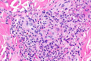
 Leukocytoclastic vasculitis. H&E stain. | |
|
| |
| LM | small vessels intramural inflammatory cells (neutrophils), vessel damage (fibrin deposition) |
| LM DDx | dermatitides with perivascular inflammation |
| Stains | PAS -ve |
| Site | blood vessels - see vasculitides |
|
| |
| Signs | palpable purpura |
| Prevalence | uncommon |
| Prognosis | dependent on underlying cause |
General
- Most common cutaneous vasculitis.[1]
Clinical:
- Palpable purpura, usu. lower extremity.
Microscopic
Features:[1]
- Small upper dermis vessels with:
- Neutrophils.
- Fragmentation of neutrophils (leukocytoclasia).
- Vessel damage: fibrin deposition (bright pink acellular stuff).
- Neutrophils.
Has a very broad DDx:[1]
- Infectious:
- Bacterial.
- Viral.
- Fungal.
- Vasculitic disorders:
- ANCA mediated vasculitides:
- Henoch–Schönlein purpura.[2]
- Urticarial vasculitis.
- Other:
- Connective tissue disease, e.g. mixed connective tissue disease, SLE, rheumatoid arthritis.
- Cryoglobulinemia - may be due to multiple myeloma, hepatitis C; have intravascular thrombi.
- Paraneoplastic.
- Drugs.
Image
Case
www
Stains
- PAS - look for fungus.
See also
References
- ↑ 1.0 1.1 1.2 Brinster, NK. (Nov 2008). "Dermatopathology for the surgical pathologist: a pattern-based approach to the diagnosis of inflammatory skin disorders (part II).". Adv Anat Pathol 15 (6): 350-69. doi:10.1097/PAP.0b013e31818b1ac6. PMID 18948765.
- ↑ Kraft, DM.; Mckee, D.; Scott, C. (Aug 1998). "Henoch-Schönlein purpura: a review.". Am Fam Physician 58 (2): 405-8, 411. PMID 9713395.