Epithelioid hemangioendothelioma
Jump to navigation
Jump to search
Epithelioid hemangioendothelioma is rare malignant vascular tumour.
| Epithelioid hemangioendothelioma | |
|---|---|
| Diagnosis in short | |
|
| |
| LM | large epithelioid perivascular cells with abundant pale eosinophilic cytoplasm and cytoplasmic vacuolation ("blister cells") - may form lumen and have RBC within, vesicular nucleus +/-prominent nucleolus; tuft-like projections into capillaries; tumour cells may be in well-circumscribed paucicellular nodules or more cellular poorly formed aggregates |
| LM DDx | epithelioid angiosarcoma, hemangioma |
| IHC | CD31 +ve, CD34 +ve, factor VIII +ve |
| Site | soft tissue - see vascular tumours |
|
| |
| Prevalence | rare |
It should not be confused with epithelioid hemangioma.
General
Microscopic
Features:[2]
- Large epithelioid perivascular cells with:
- Abundant pale eosinophilic cytoplasm.
- Cytoplasmic vacuolation (some cells) - AKA "blister cells" - key feature.
- May form lumen and have RBC within.
- Vesicular nucleus with prominent nucleolus in some cells.
- Tuft-like projections into capillaries.
- Tumour cells may be in well-circumscribed paucicellular nodules or more cellular poorly formed aggregates.
DDx:
- Angiosarcoma, epithelioid.
- Hemangioma.
Images
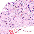
Epithelioid hemangioendothelioma. (WC)

www:
IHC
Features:[2]
- CD31 +ve.
- CD34 +ve.
- Factor VIII +ve.
See also
References
- ↑ Humphrey, Peter A; Dehner, Louis P; Pfeifer, John D (2008). The Washington Manual of Surgical Pathology (1st ed.). Lippincott Williams & Wilkins. pp. 603. ISBN 978-0781765275.
- ↑ 2.0 2.1 2.2 Gupta, R.; Mathur, SR.; Gupta, SD.; Durgapal, P.; Iyer, VK.; Das, CJ.; Shalimar, SK.; Acharya, . (2010). "Hepatic epithelioid hemangioendothelioma: A diagnostic pitfall in aspiration cytology.". Cytojournal 6: 25. doi:10.4103/1742-6413.58951. PMID 20165548.