Keratocystic odontogenic tumour
Jump to navigation
Jump to search
Keratocystic odontogenic tumour, abbreviated KOT, is an uncommon odontogenic tumour.
It was rreviously known as odontogenic keratocyst, abbreviated OKC.[1]
General
- May be associated with nevoid basal cell carcinoma syndrome.
Clinical
Features:[2]
- Most common presentation: swelling.
Gross
- Location: usually mandible.
- May mimic ameloblastoma radiologically.
Microscopic
Features: [3]
- Stratified epithelium (resembling squamous epithelium) with:
- "Ribbon-like appearance" - important.
- Typically 8-10 cell layers thick - with relatively uniform thickness.
- Lacks rete ridges.
- Palisaded basal cell layer.
- "Ribbon-like appearance" - important.
- Parakeratosis (keratinized cells with nuclei) - key feature.
- Artefactual separation of epithelium from the basement membrane.
DDx:
- Odontogenic cyst.
- Orthokeratinized odontogenic cyst[4] - usu. dentigerous cyst - has orthokeratosis instead of parakeratosis.
- Orthokeratosis = keratinized cells no nuclei; parakeratosis = keratinized cell with nuclei.
- Orthokeratinized odontogenic cyst[4] - usu. dentigerous cyst - has orthokeratosis instead of parakeratosis.
Images
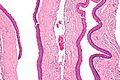
KOT - intermed. mag. (WC)
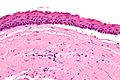
KOT - very high mag. (WC)
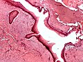
KOT - poor quality. (WC)
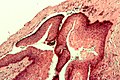
KOT - showing artefactual clefting - poor quality. (WC)


www:
See also
References
- ↑ Madras, J.; Lapointe, H. (Mar 2008). "Keratocystic odontogenic tumour: reclassification of the odontogenic keratocyst from cyst to tumour.". J Can Dent Assoc 74 (2): 165-165h. PMID 18353202.
- ↑ Habibi, A.; Saghravanian, N.; Habibi, M.; Mellati, E.; Habibi, M. (Sep 2007). "Keratocystic odontogenic tumor: a 10-year retrospective study of 83 cases in an Iranian population.". J Oral Sci 49 (3): 229-35. PMID 17928730.
- ↑ Thompson LDR. Head and neck pathology - (Foundations in diagnostic pathology). Goldblum JR, Ed.. Churchill Livingstone. 2006. ISBN 0-443-06960-3.
- ↑ Macdonald-Jankowski, DS. (Dec 2010). "Orthokeratinized odontogenic cyst: a systematic review.". Dentomaxillofac Radiol 39 (8): 455-67. doi:10.1259/dmfr/19728573.
- ↑ URL: http://www.sciencedirect.com/science/article/pii/S0968605305000992#fig5. Accessed on: 11 March 2013.