Metaphyseal fibrous defect
Jump to navigation
Jump to search
| Template:Metaphyseal fibrous defect | |
|---|---|
| Diagnosis in short | |
|
| |
| Synonyms | Nonossifying fibroma |
| Clinical history | Incidental radiograhic finding |
| Radiology | Lucent defect |
General
- Common
- Non-neoplastic
- Self-limited
- Skeletally immature individuals, children and adolescent
- Often small lesions discovered an radiographic incidentaloma
Synonyms
- Nonossifying fibroma (larger but otherwise identical)
- Fibrous cortical defect
- Fibrous metaphyseal defect
- Fibroxanthoma of bone
Site
- Metaphysis of distal femur or proximal tibia (80%)
- Cortical
- Metaphysis
- Long bones
- Eccentric location
Gross
Firm, granular, brown to yellow to red
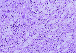
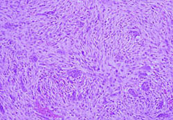
Microscopic
Spindle cells without cytologic atypia are arranged in a storiform pattern with scattered chronic inflammatory cells and benign giant cells. Foam cells and hemosiderin deposition are present. Mitoses are seen but cytologic atypia is absent.
Differential Diagnosis
- Giant cell tumor of bone (epiphyseal location, occurs in adults)
Relevant Diagnostic Groups
- FOG MACHINES - acronym for radiographically lytic bone lesions <ref {{http://radiopaedia.org/articles/lytic-bone-lesion-mnemonic}} /ref>
- Giant cell lesions of bone.
- Spindle cell lesions of bone.
Images
- http://njms2.umdnj.edu/tutorweb/casegifs/nofhe1.jpg
- http://njms2.umdnj.edu/tutorweb/casegifs/nofhe2.jpg
- http://njms2.umdnj.edu/tutorweb/casegifs/nofhe1.jpg
- http://pathologyoutlines.com/wick/non-ossifying%20fibroma%20(metaphyseal%20fibrous%20defect)%20micro.jpg
%20micro.jpg)
Stains
Not relevant.
IHC
Not relevant
Molecular
Not relevant
Syndromes
Jaffe-Campanacci syndrome <refTemplate:Http://www.bonetumor.org/plasma-cell-tumors/jaffe-campanacci-syndrome/ref>
Clinical history
- Incidental radiographic finding
- Pathologic fracture
Radiographic findings
Sharply demarcated, lucent, loculated, meta-diaphyseal lesion surrounded by a rim of sclerotic bone
Sign out
BONE; CURETTAGE: METAPHYSEAL FIBROUS DEFECT.
See also
- http://njms2.umdnj.edu/tutorweb/case8.htm
- http://www.pathologyoutlines.com/topic/bonemetaphysealfibrousdefect.html
- http://radiopaedia.org/articles/fibrous-cortical-defect
- http://radiopaedia.org/articles/non-ossifying-fibroma-1