Coffee bean nucleus
Jump to navigation
Jump to search

File:Coffee Beans macro 1.jpg
Coffee beans.
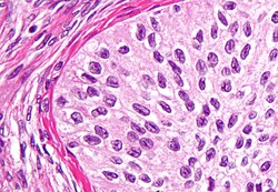
Coffee bean nucleus is a cell nucleus that looks like a coffee bean. Coffee bean nuclei have a limited differential diagnosis.
Microscopic
- Coffee bean nuclei are ellipsoid and have a central groove.
Images
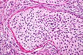
Coffee bean nuclei in a Brenner tumour (WC)

Classic differential diagnosis
Others considerations
- Invasive ductal carcinoma of the pancreas.
- Langerhans cell histiocytosis.
- Papillary thyroid carcinoma. (???)
See also
References
- ↑ Vodovnik, A. (Jun 2002). "Bladder-washing cytology of metastatic ovarian granulosa cell tumor.". Diagn Cytopathol 26 (6): 387-8. doi:10.1002/dc.10095. PMID 12112830.
- ↑ Ahr, A.; Arnold, G.; Göhring, UJ.; Costa, S.; Scharl, A.; Gauwerky, JF.. "Cytology of ascitic fluid in a patient with metastasizing malignant Brenner tumor of the ovary. A case report.". Acta Cytol 41 (4 Suppl): 1299-304. PMID 9990262.