Difference between revisions of "Pseudoepitheliomatous hyperplasia"
Jump to navigation
Jump to search
(more) |
|||
| Line 6: | Line 6: | ||
#[[Granular cell tumour]]. | #[[Granular cell tumour]]. | ||
#Adjacent to an ulcer. | #Adjacent to an ulcer. | ||
===Microscopic=== | |||
Features: | |||
*Epidermal thickening.<ref name=pmid21399447>{{Cite journal | last1 = Zayour | first1 = M. | last2 = Lazova | first2 = R. | title = Pseudoepitheliomatous hyperplasia: a review. | journal = Am J Dermatopathol | volume = 33 | issue = 2 | pages = 112-22; quiz 123-6 | month = Apr | year = 2011 | doi = 10.1097/DAD.0b013e3181fcfb47 | PMID = 21399447 }}</ref> | |||
Images: | Images: | ||
Revision as of 19:19, 10 November 2012
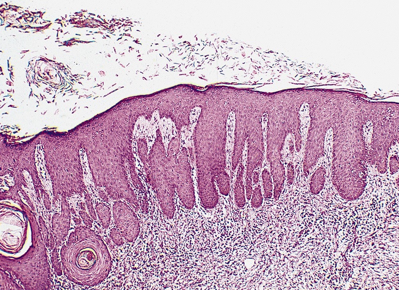
Pseudoepitheliomatous hyperplasia is mimic of squamous cell carcinoma.
It is seen in:
- Fungal infections.
- Inflammatory papillary hyperplasia.
- Granular cell tumour.
- Adjacent to an ulcer.
Microscopic
Features:
- Epidermal thickening.[1]
Images:
- Pseudoepitheliomatous hyperplasia (pathologyoutlines.com).[2]
- Pseudoepitheliomatous hyperplasia (the-dermatologist.com).[3]
References
- ↑ Zayour, M.; Lazova, R. (Apr 2011). "Pseudoepitheliomatous hyperplasia: a review.". Am J Dermatopathol 33 (2): 112-22; quiz 123-6. doi:10.1097/DAD.0b013e3181fcfb47. PMID 21399447.
- ↑ URL: http://www.pathologyoutlines.com/topic/penscrotumpenssqhyper.html. Accessed on: 10 November 2012.
- ↑ URL: http://www.the-dermatologist.com/content/treating-rare-fungal-infections-coccidioidomycosis. Accessed on: 10 November 2012.