Difference between revisions of "Nodular fasciitis"
Jump to navigation
Jump to search
(tweak) |
(more) |
||
| Line 8: | Line 8: | ||
| LMDDx = | | LMDDx = | ||
| Stains = | | Stains = | ||
| IHC = | | IHC = CD34 -ve, desmin -ve, SMA -ve, S-100 -ve, AE1/AE3 -ve. | ||
| EM = | | EM = | ||
| Molecular = | | Molecular = t(15;15) ? | ||
| IF = | | IF = | ||
| Gross = | | Gross = | ||
| Line 29: | Line 29: | ||
}} | }} | ||
'''Nodular fasciitis''' is an uncommon [[soft tissue lesion]]. It should '''not''' to be confused with [[necrotizing fasciitis]]. | '''Nodular fasciitis''' is an uncommon [[soft tissue lesion]]. It should '''not''' to be confused with [[necrotizing fasciitis]]. | ||
==General== | |||
*Benign. | *Benign. | ||
*All age groups. | *All age groups. | ||
*Associated with trauma. | *Associated with trauma. | ||
==Microscopic== | |||
Features:<ref name=Ref_WMSP606>{{Ref WMSP|606}}</ref><ref>{{cite journal |author=de Feraudy S, Fletcher CD |title=Intradermal nodular fasciitis: a rare lesion analyzed in a series of 24 cases |journal=Am. J. Surg. Pathol. |volume=34 |issue=9 |pages=1377–81 |year=2010 |month=September |pmid=20716998 |doi=10.1097/PAS.0b013e3181ed7374 |url=}}</ref> | Features:<ref name=Ref_WMSP606>{{Ref WMSP|606}}</ref><ref>{{cite journal |author=de Feraudy S, Fletcher CD |title=Intradermal nodular fasciitis: a rare lesion analyzed in a series of 24 cases |journal=Am. J. Surg. Pathol. |volume=34 |issue=9 |pages=1377–81 |year=2010 |month=September |pmid=20716998 |doi=10.1097/PAS.0b013e3181ed7374 |url=}}</ref> | ||
*Usu. well-circumscribed. | *Usu. well-circumscribed. | ||
| Line 73: | Line 73: | ||
www: | www: | ||
*[http://www.humpath.com/nodular-fasciitis NF (humpath.com)]. | *[http://www.humpath.com/nodular-fasciitis NF (humpath.com)]. | ||
==IHC== | |||
Routine spindle cell panel: | Routine spindle cell panel: | ||
*CD34 -ve. | *CD34 -ve. | ||
*Desmin -ve. | *Desmin -ve. | ||
*SMA -ve. | *SMA -ve. | ||
* | *S-100 -ve. | ||
*AE1/AE3 -ve. | *AE1/AE3 -ve. | ||
| Line 86: | Line 86: | ||
*Vimentin +ve. | *Vimentin +ve. | ||
==Molecular== | |||
*Evolving - case reports. | *Evolving - case reports. | ||
**t(15;15)(q13;q25).<ref name=pmid12606136>{{cite journal |author=Velagaleti GV, Tapper JK, Panova NE, Miettinen M, Gatalica Z |title=Cytogenetic findings in a case of nodular fasciitis of subclavicular region |journal=Cancer Genet. Cytogenet. |volume=141 |issue=2 |pages=160–3 |year=2003 |month=March |pmid=12606136 |doi= |url=}}</ref> | **t(15;15)(q13;q25).<ref name=pmid12606136>{{cite journal |author=Velagaleti GV, Tapper JK, Panova NE, Miettinen M, Gatalica Z |title=Cytogenetic findings in a case of nodular fasciitis of subclavicular region |journal=Cancer Genet. Cytogenet. |volume=141 |issue=2 |pages=160–3 |year=2003 |month=March |pmid=12606136 |doi= |url=}}</ref> | ||
Revision as of 03:05, 29 September 2013
| Nodular fasciitis | |
|---|---|
| Diagnosis in short | |
|
Template:Px Nodular fasciitis. H&E stain. | |
|
| |
| LM | usu. well-circumscribed, clusters of (non-pleomorphic) spindle cells, inflammation (lymphocytes), microcysts in cellular regions - uncommon, mitoses - common, extravasated RBCs. |
| IHC | CD34 -ve, desmin -ve, SMA -ve, S-100 -ve, AE1/AE3 -ve. |
| Molecular | t(15;15) ? |
| Site | soft tissue - fibroblastic/myofibroblastic tumours |
|
| |
| Clinical history | associated with trauma |
| Prevalence | uncommon |
| Prognosis | benign |
Nodular fasciitis is an uncommon soft tissue lesion. It should not to be confused with necrotizing fasciitis.
General
- Benign.
- All age groups.
- Associated with trauma.
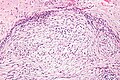
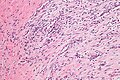
Microscopic
- Usu. well-circumscribed.
- Clusters of (non-pleomorphic) spindle cells.
- Inflammation (lymphocytes).
- Microcysts in cellular regions - uncommon - discriminatory.
- Mitoses - common.
- Extravasated RBCs.
- Tissue culture-like/CNS-like morphology.
- Thick (keloid-like) collagen bundles - key feature.
- Extravasated RBCs.
- Inflammation.
- +/-Giant cells.
Notes:
- No significant nuclear atypia.
- No atypical mitoses.
- May be cellular.
DDx:[5]
- Myxoid DFSP.
- Cellular dermatofibroma.
- Desmoid-type fibromatosis.
Images
- Nodular fasciitis - intermed mag.jpg
NF - case 1 - intermed. mag. (WC)
NF - case 1 - high mag. (WC)
- Nodular fasciitis -2- intermed mag.jpg
NF - case 2 - intermed. mag. (WC)
NF - case 2 - high mag. (WC)


- Nodular fasciitis (1).JPG
NF - low mag. (WC)
- Nodular fasciitis (2).JPG
NF - high mag. (WC)
www:
IHC
Routine spindle cell panel:
- CD34 -ve.
- Desmin -ve.
- SMA -ve.
- S-100 -ve.
- AE1/AE3 -ve.
Others:
- H-caldesmon -ve.
- EMA -ve.
- Vimentin +ve.
Molecular
- Evolving - case reports.
- t(15;15)(q13;q25).[6]
See also
References
- ↑ Humphrey, Peter A; Dehner, Louis P; Pfeifer, John D (2008). The Washington Manual of Surgical Pathology (1st ed.). Lippincott Williams & Wilkins. pp. 606. ISBN 978-0781765275.
- ↑ de Feraudy S, Fletcher CD (September 2010). "Intradermal nodular fasciitis: a rare lesion analyzed in a series of 24 cases". Am. J. Surg. Pathol. 34 (9): 1377–81. doi:10.1097/PAS.0b013e3181ed7374. PMID 20716998.
- ↑ Dickson, B. 26 April 2011.
- ↑ URL: http://anvita.info/wiki/Nodular_Fasciitis. Accessed on: 11 November 2011.
- ↑ URL: http://www.mckeedermpath.com/SPOT%20DIAGNOSIS%20CASE%20268.html. Accessed on: 11 November 2011.
- ↑ Velagaleti GV, Tapper JK, Panova NE, Miettinen M, Gatalica Z (March 2003). "Cytogenetic findings in a case of nodular fasciitis of subclavicular region". Cancer Genet. Cytogenet. 141 (2): 160–3. PMID 12606136.