Difference between revisions of "Cardiac myxoma"
(redirect) |
(→Images) |
||
| (7 intermediate revisions by the same user not shown) | |||
| Line 1: | Line 1: | ||
{{ Infobox diagnosis | |||
| Name = {{PAGENAME}} | |||
| Image = Atrial_myxoma_edge_high_mag.jpg | |||
| Width = | |||
| Caption = Atrial myxoma. [[H&E stain]]. | |||
| Micro = myxoid material (extra cellular), myxoma cells (stellate, polygonal or spindled morphology; +/-multinucleated, inconspicuous [[nucleoli]]; abundant cytoplasm), calcified elastic fibers (''gamna bodies''), +/-hemorrhage, +/-endothelial covering, +/-ossification, +/-fibrosis | |||
| Subtypes = | |||
| LMDDx = [[myxofibrosarcoma]], [[myxoid liposarcoma]], [[papillary fibroelastoma]] | |||
| Stains = | |||
| IHC = | |||
| EM = | |||
| Molecular = | |||
| IF = | |||
| Gross = usu. atrium, usu. left side, classically projects into the lumen | |||
| Grossing = | |||
| Site = [[heart]] | |||
| Assdx = | |||
| Syndromes = | |||
| Clinicalhx = | |||
| Signs = | |||
| Symptoms = +/-dyspnea, +/-neurologic symptoms | |||
| Prevalence = most common heart tumour, generally uncommon | |||
| Bloodwork = | |||
| Rads = | |||
| Endoscopy = | |||
| Prognosis = | |||
| Other = | |||
| ClinDDx = other heart tumours | |||
}} | |||
{{ Infobox external links | |||
| Name = {{PAGENAME}} | |||
| EHVSC = | |||
| EHVSC_mult = | |||
| pathprotocols = | |||
| wikipedia = atrial myxoma | |||
| pathoutlines = | |||
| rosaicollection = 1267 | |||
}} | |||
'''Cardiac myxoma''' is the most common [[Cardiac tumours|tumour of the heart]]. They are typical in the atrium; thus, a synonym is '''atrial myxoma'''. | |||
==General== | |||
*Uncommon. | |||
*Clinical: may lead to cerebral infarction.<ref name=pmid3188128>{{cite journal |author=Knepper LE, Biller J, Adams HP, Bruno A |title=Neurologic manifestations of atrial myxoma. A 12-year experience and review |journal=Stroke |volume=19 |issue=11 |pages=1435-40 |year=1988 |month=November |pmid=3188128 |doi= |url=http://stroke.ahajournals.org/cgi/reprint/19/11/1435}}</ref> | |||
*Diagnosed by imaging. | |||
*May be familial, i.e. [[Carney complex]] ([[AKA]] NAME syndrome, [[AKA]] LAMB syndrome).<ref>{{Ref WMSP|135}}</ref> | |||
**NAME = Nevi, Atrial myxoma, Myxoid neurofibroma, and Ephelides (freckles<ref>URL: [http://emedicine.medscape.com/article/1119293-overview http://emedicine.medscape.com/article/1119293-overview]. Accessed on: 7 January 2011.</ref>). | |||
**LAMB = Lentigines, Atrial myxomas, Mucocutaneous myxomas, Blue nevi. | |||
Most common presentations:<ref name=pmid3188128/> | |||
*[[Dyspnea]] - 45%. | |||
*Neurologic symptoms 36%. | |||
==Gross== | |||
Location:<ref name=pmid3188128/> | |||
*Usually atrial. | |||
*Usually left side ~60%.<ref name=pmid12006696/> | |||
Features:<ref name=pmid12006696>{{cite journal |author=Grebenc ML, Rosado-de-Christenson ML, Green CE, Burke AP, Galvin JR |title=Cardiac myxoma: imaging features in 83 patients |journal=Radiographics |volume=22 |issue=3 |pages=673-89 |year=2002 |pmid=12006696 |doi= |url=http://radiographics.rsna.org/content/22/3/673.long}}</ref> | |||
*Lobular surface. | |||
*Smooth surface. | |||
===Image=== | |||
<gallery> | |||
Image:Myxoma.jpg |Cardiac myxoma. (WC/AFIP) | |||
</gallery> | |||
==Microscopic== | |||
Features:<ref name=pmid12006696/> | |||
*[[myxoid stroma|Myxoid]] material - extra cellular - '''key feature'''. | |||
*Myxoma cells:<ref name=pmid16508920>{{Cite journal | last1 = Orlandi | first1 = A. | last2 = Ciucci | first2 = A. | last3 = Ferlosio | first3 = A. | last4 = Genta | first4 = R. | last5 = Spagnoli | first5 = LG. | last6 = Gabbiani | first6 = G. | title = Cardiac myxoma cells exhibit embryonic endocardial stem cell features. | journal = J Pathol | volume = 209 | issue = 2 | pages = 231-9 | month = Jun | year = 2006 | doi = 10.1002/path.1959 | PMID = 16508920 }}</ref> | |||
**Stellate, polygonal or spindled morphology. | |||
**+/-Multinucleated. | |||
**Inconspicuous [[nucleoli]]. | |||
**Abundant cytoplasm. | |||
*Calcified elastic fibers - ''gamna bodies''. | |||
*Hemorrhage common. | |||
*Often covered by endothelium. | |||
*+/-Ossification. | |||
*+/-Fibrosis. | |||
DDx:<ref>{{Ref DCHH|79}}</ref> | |||
*[[Papillary fibroelastoma]] - not really in the histomorphologic [[differential diagnosis]]. | |||
*[[Myxofibrosarcoma]] - nuclear pleomorphism. | |||
*[[Myxoid liposarcoma]] - chickenwire vasculature. | |||
===Images=== | |||
<gallery> | |||
Image:Atrial myxoma low mag.jpg| AM - low mag. | |||
Image:Atrial myxoma intermed mag.jpg| AM - intermed. mag. | |||
Image:Atrial myxoma high mag.jpg| AM - high mag. | |||
Image:Atrial myxoma edge low mag.jpg| AM with interface to muscular wall - low mag. | |||
Image:Atrial myxoma edge high mag.jpg| AM with endothelium - high mag. | |||
</gallery> | |||
====www==== | |||
*[http://radiographics.rsna.org/content/22/3/673/F3.expansion.html Atrial myxoma (rsna.org)].<ref name=pmid12006696/> | |||
*[http://radiographics.rsna.org/content/22/3/673/F4.expansion.html Gamna bodies (rsna.org)].<ref name=pmid12006696/> | |||
==Sign out== | |||
<pre> | |||
MASS, LEFT ATRIUM, EXCISION: | |||
- MYXOMA. | |||
</pre> | |||
===Micro=== | |||
The sections show paucicellular myxoid material containing polygonal and spindled cells with eosinophilic myxoid cytoplasm, bland nuclei, inconspicuous nucleoli and focal multinucleation (myxoma cells). Hemosiderin-laden macrophages, calcified elastic fibres (gamna bodies) and scattered inflammatory cells are also present. There is no nuclear atypia. Mitotic activity is not evident. Several sections show fresh hemorrhage. The edge has a fibrotic rim and appears to be covered by endothelium. No cardiac muscle is identified. | |||
==See also== | |||
*[[Cardiac tumours]]. | |||
*[[Myxoid lesions]]. | |||
==References== | |||
{{Reflist|2}} | |||
[[Category:Cardiovascular pathology]] | |||
[[Category:Cardiac tumours]] | |||
Latest revision as of 16:11, 23 July 2016
| Cardiac myxoma | |
|---|---|
| Diagnosis in short | |
 Atrial myxoma. H&E stain. | |
|
| |
| LM | myxoid material (extra cellular), myxoma cells (stellate, polygonal or spindled morphology; +/-multinucleated, inconspicuous nucleoli; abundant cytoplasm), calcified elastic fibers (gamna bodies), +/-hemorrhage, +/-endothelial covering, +/-ossification, +/-fibrosis |
| LM DDx | myxofibrosarcoma, myxoid liposarcoma, papillary fibroelastoma |
| Gross | usu. atrium, usu. left side, classically projects into the lumen |
| Site | heart |
|
| |
| Symptoms | +/-dyspnea, +/-neurologic symptoms |
| Prevalence | most common heart tumour, generally uncommon |
| Clin. DDx | other heart tumours |
| Cardiac myxoma | |
|---|---|
| External resources | |
| Wikipedia | atrial myxoma |
| Rosai Collection | 1267 |
Cardiac myxoma is the most common tumour of the heart. They are typical in the atrium; thus, a synonym is atrial myxoma.
General
- Uncommon.
- Clinical: may lead to cerebral infarction.[1]
- Diagnosed by imaging.
- May be familial, i.e. Carney complex (AKA NAME syndrome, AKA LAMB syndrome).[2]
- NAME = Nevi, Atrial myxoma, Myxoid neurofibroma, and Ephelides (freckles[3]).
- LAMB = Lentigines, Atrial myxomas, Mucocutaneous myxomas, Blue nevi.
Most common presentations:[1]
- Dyspnea - 45%.
- Neurologic symptoms 36%.
Gross
Location:[1]
- Usually atrial.
- Usually left side ~60%.[4]
Features:[4]
- Lobular surface.
- Smooth surface.
Image
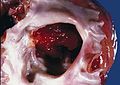
Cardiac myxoma. (WC/AFIP)

Microscopic
Features:[4]
- Myxoid material - extra cellular - key feature.
- Myxoma cells:[5]
- Stellate, polygonal or spindled morphology.
- +/-Multinucleated.
- Inconspicuous nucleoli.
- Abundant cytoplasm.
- Calcified elastic fibers - gamna bodies.
- Hemorrhage common.
- Often covered by endothelium.
- +/-Ossification.
- +/-Fibrosis.
DDx:[6]
- Papillary fibroelastoma - not really in the histomorphologic differential diagnosis.
- Myxofibrosarcoma - nuclear pleomorphism.
- Myxoid liposarcoma - chickenwire vasculature.
Images
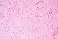
AM - low mag.
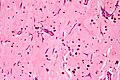
AM - intermed. mag.
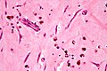
AM - high mag.
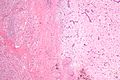
AM with interface to muscular wall - low mag.
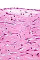
AM with endothelium - high mag.


www
Sign out
MASS, LEFT ATRIUM, EXCISION:
- MYXOMA.
Micro
The sections show paucicellular myxoid material containing polygonal and spindled cells with eosinophilic myxoid cytoplasm, bland nuclei, inconspicuous nucleoli and focal multinucleation (myxoma cells). Hemosiderin-laden macrophages, calcified elastic fibres (gamna bodies) and scattered inflammatory cells are also present. There is no nuclear atypia. Mitotic activity is not evident. Several sections show fresh hemorrhage. The edge has a fibrotic rim and appears to be covered by endothelium. No cardiac muscle is identified.
See also
References
- ↑ 1.0 1.1 1.2 Knepper LE, Biller J, Adams HP, Bruno A (November 1988). "Neurologic manifestations of atrial myxoma. A 12-year experience and review". Stroke 19 (11): 1435-40. PMID 3188128. http://stroke.ahajournals.org/cgi/reprint/19/11/1435.
- ↑ Humphrey, Peter A; Dehner, Louis P; Pfeifer, John D (2008). The Washington Manual of Surgical Pathology (1st ed.). Lippincott Williams & Wilkins. pp. 135. ISBN 978-0781765275.
- ↑ URL: http://emedicine.medscape.com/article/1119293-overview. Accessed on: 7 January 2011.
- ↑ 4.0 4.1 4.2 4.3 4.4 Grebenc ML, Rosado-de-Christenson ML, Green CE, Burke AP, Galvin JR (2002). "Cardiac myxoma: imaging features in 83 patients". Radiographics 22 (3): 673-89. PMID 12006696. http://radiographics.rsna.org/content/22/3/673.long.
- ↑ Orlandi, A.; Ciucci, A.; Ferlosio, A.; Genta, R.; Spagnoli, LG.; Gabbiani, G. (Jun 2006). "Cardiac myxoma cells exhibit embryonic endocardial stem cell features.". J Pathol 209 (2): 231-9. doi:10.1002/path.1959. PMID 16508920.
- ↑ Tadrous, Paul.J. Diagnostic Criteria Handbook in Histopathology: A Surgical Pathology Vade Mecum (1st ed.). Wiley. pp. 79. ISBN 978-0470519035.