Difference between revisions of "Follicular dendritic cell sarcoma"
Jump to navigation
Jump to search
(Teeak) |
|||
| (15 intermediate revisions by the same user not shown) | |||
| Line 1: | Line 1: | ||
{{ Infobox diagnosis | {{ Infobox diagnosis | ||
| Name = {{PAGENAME}} | | Name = {{PAGENAME}} | ||
| Image = | | Image = Follicular dendritic cell sarcoma - alt -- very high mag.jpg | ||
| Width = | | Width = | ||
| Caption = | | Caption = [[Micrograph]] showing a follicular dendritic cell sarcoma. The (benign) interspersed lymphocytes are a characteristic finding. [[H&E stain]]. | ||
| Synonyms = | | Synonyms = follicular dendritic cell tumour, follicular dendritic cell neoplasm | ||
| Micro = oval or spindle-shaped cellular & nuclear morphology, variable architecture (sheets, fascicles, whorles, storiform pattern), nuclei with small nucleoli and clear or dispersed chromatin, multinucleated cells, interspersed small lymphocytes, +/-necrosis, +/-marked nuclear atypia, +/-abundant mitoses. | | Micro = oval or spindle-shaped cellular & nuclear morphology, variable architecture (sheets, fascicles, whorles, storiform pattern), nuclei with small nucleoli and clear or dispersed chromatin, multinucleated cells, interspersed small lymphocytes, +/-necrosis, +/-marked nuclear atypia, +/-abundant mitoses. | ||
| Subtypes = | | Subtypes = | ||
| LMDDx = [[histiocytic sarcoma]], [[thymoma]], other [[spindle cell lesions]] | | LMDDx = [[histiocytic sarcoma]], [[thymoma]], other [[spindle cell lesions]] | ||
| Stains = | | Stains = | ||
| IHC = CD21 +ve, CD35 +ve, Ki-M4p +ve, Ki-FDRC1p +ve, vimentin +ve | | IHC = CD21 +ve, CD23 +ve, CD35 +ve, Ki-M4p +ve, Ki-FDRC1p +ve, vimentin +ve | ||
| EM = | | EM = | ||
| Molecular = | | Molecular = | ||
| Line 17: | Line 17: | ||
| Staging = | | Staging = | ||
| Site = | | Site = | ||
| Assdx = | | Assdx = [[Castleman disease|Castleman disease, hyaline-vascular type]] | ||
| Syndromes = | | Syndromes = | ||
| Clinicalhx = | | Clinicalhx = | ||
| Line 33: | Line 33: | ||
'''Follicular dendritic cell sarcoma''', abbreviated '''FDCS''', is a very rare [[malignant]] tumour. | '''Follicular dendritic cell sarcoma''', abbreviated '''FDCS''', is a very rare [[malignant]] tumour. | ||
It is also known as '''follicular dendritic cell tumour''' (abbreviated '''FDCT''') | It is also known as '''follicular dendritic cell tumour''' (abbreviated '''FDCT'''),<ref name=pmid17515347>{{Cite journal | last1 = Leipsic | first1 = JA. | last2 = McAdams | first2 = HP. | last3 = Sporn | first3 = TA. | title = Follicular dendritic cell sarcoma of the mediastinum. | journal = AJR Am J Roentgenol | volume = 188 | issue = 6 | pages = W554-6 | month = Jun | year = 2007 | doi = 10.2214/AJR.04.1530 | PMID = 17515347 }}</ref> and '''follicular dendritic cell neoplasm'''.<ref name=pmid19391666>{{Cite journal | last1 = Denning | first1 = KL. | last2 = Olson | first2 = PR. | last3 = Maley | first3 = RH. | last4 = Flati | first4 = VR. | last5 = Myers | first5 = JL. | last6 = Silverman | first6 = JF. | title = Primary pulmonary follicular dendritic cell neoplasm: a case report and review of the literature. | journal = Arch Pathol Lab Med | volume = 133 | issue = 4 | pages = 643-7 | month = Apr | year = 2009 | doi = 10.1043/1543-2165-133.4.643 | PMID = 19391666 }}</ref> | ||
==General== | ==General== | ||
| Line 41: | Line 39: | ||
*Behave like a low-grade sarcoma.<ref name=pmid9606805>{{Cite journal | last1 = Perez-Ordoñez | first1 = B. | last2 = Rosai | first2 = J. | title = Follicular dendritic cell tumor: review of the entity. | journal = Semin Diagn Pathol | volume = 15 | issue = 2 | pages = 144-54 | month = May | year = 1998 | doi = | PMID = 9606805 }}</ref> | *Behave like a low-grade sarcoma.<ref name=pmid9606805>{{Cite journal | last1 = Perez-Ordoñez | first1 = B. | last2 = Rosai | first2 = J. | title = Follicular dendritic cell tumor: review of the entity. | journal = Semin Diagn Pathol | volume = 15 | issue = 2 | pages = 144-54 | month = May | year = 1998 | doi = | PMID = 9606805 }}</ref> | ||
*Reported association with [[Castleman disease]] (hyaline-vascular type).<ref name=pmid9606805/> | *Reported association with [[Castleman disease]] (hyaline-vascular type).<ref name=pmid9606805/> | ||
WHO 2001 classification of dendritic cell neoplasms:<ref name=pmid17636477>{{Cite journal | last1 = Kairouz | first1 = S. | last2 = Hashash | first2 = J. | last3 = Kabbara | first3 = W. | last4 = McHayleh | first4 = W. | last5 = Tabbara | first5 = IA. | title = Dendritic cell neoplasms: an overview. | journal = Am J Hematol | volume = 82 | issue = 10 | pages = 924-8 | month = Oct | year = 2007 | doi = 10.1002/ajh.20857 | PMID = 17636477 }}</ref> | |||
*[[Langerhans cell histiocytosis]]. | |||
*Langerhans cell sarcoma. | |||
*Interdigitating dendritic cell sarcoma/tumour. | |||
*Follicular dendritic cell sarcoma/tumour. | |||
*Dendritic cell sarcoma, not specified otherwise. | |||
==Gross== | |||
Features: | |||
*Often a head and neck lesion.{{fact}} | |||
==Microscopic== | ==Microscopic== | ||
| Line 58: | Line 67: | ||
*[[Histiocytic sarcoma]].<ref name=pmid17324277>{{Cite journal | last1 = Alexiev | first1 = BA. | last2 = Sailey | first2 = CJ. | last3 = McClure | first3 = SA. | last4 = Ord | first4 = RA. | last5 = Zhao | first5 = XF. | last6 = Papadimitriou | first6 = JC. | title = Primary histiocytic sarcoma arising in the head and neck with predominant spindle cell component. | journal = Diagn Pathol | volume = 2 | issue = | pages = 7 | month = | year = 2007 | doi = 10.1186/1746-1596-2-7 | PMID = 17324277 | url = http://www.diagnosticpathology.org/content/2/1/7 }}</ref> | *[[Histiocytic sarcoma]].<ref name=pmid17324277>{{Cite journal | last1 = Alexiev | first1 = BA. | last2 = Sailey | first2 = CJ. | last3 = McClure | first3 = SA. | last4 = Ord | first4 = RA. | last5 = Zhao | first5 = XF. | last6 = Papadimitriou | first6 = JC. | title = Primary histiocytic sarcoma arising in the head and neck with predominant spindle cell component. | journal = Diagn Pathol | volume = 2 | issue = | pages = 7 | month = | year = 2007 | doi = 10.1186/1746-1596-2-7 | PMID = 17324277 | url = http://www.diagnosticpathology.org/content/2/1/7 }}</ref> | ||
*[[Thymoma]]. | *[[Thymoma]]. | ||
*[[Spindle cell lesions]]. | *[[Spindle cell lesions]] such as [[sarcomatoid carcinoma]]. | ||
*[[Meningioma]].{{fact}} | |||
===Images=== | ===Images=== | ||
==Related images== | |||
<gallery> | |||
Image: Follicular dendritic cell sarcoma -- very low mag.jpg | FDCS - very low mag. (WC) | |||
Image: Follicular dendritic cell sarcoma -- low mag.jpg | FDCS - low mag. (WC) | |||
Image: Follicular dendritic cell sarcoma - alt -- low mag.jpg | FDCS - low mag. (WC) | |||
Image: Follicular dendritic cell sarcoma -- intermed mag.jpg | FDCS - intermed. mag. (WC) | |||
Image: Follicular dendritic cell sarcoma -- high mag.jpg | FDCS - high mag. (WC) | |||
Image: Follicular dendritic cell sarcoma -- very high mag.jpg | FDCS - very high mag. (WC) | |||
Image: Follicular dendritic cell sarcoma - alt -- very high mag.jpg | FDCS - very high mag. (WC) | |||
</gallery> | |||
====www==== | |||
*[http://www.ajronline.org/doi/full/10.2214/AJR.04.1530 FDCS (ajronline.org)].<ref name=pmid17515347/> | *[http://www.ajronline.org/doi/full/10.2214/AJR.04.1530 FDCS (ajronline.org)].<ref name=pmid17515347/> | ||
*[http://www.npplweb.com/manuscripts/2MXAJX4VWD/0/imgfolder/WJSMRO_2_1_Fig2.jpg FDCS (npplweb.com)].<ref>URL: [http://www.npplweb.com/wjsmro/fulltext/2/1 http://www.npplweb.com/wjsmro/fulltext/2/1]. Accessed on: September 13, 2014.</ref> | *[http://www.npplweb.com/manuscripts/2MXAJX4VWD/0/imgfolder/WJSMRO_2_1_Fig2.jpg FDCS (npplweb.com)].<ref>URL: [http://www.npplweb.com/wjsmro/fulltext/2/1 http://www.npplweb.com/wjsmro/fulltext/2/1]. Accessed on: September 13, 2014.</ref> | ||
*[http://path.upmc.edu/cases/case55/dx.html FDCT - several crappy images (upmc.edu)]. | *[http://path.upmc.edu/cases/case55/dx.html FDCT - several crappy images (upmc.edu)]. | ||
| Line 78: | Line 99: | ||
*Muscle-specific actin +ve/-ve. | *Muscle-specific actin +ve/-ve. | ||
*EMA +ve/-ve. | *EMA +ve/-ve. | ||
Additional stains:<ref name=pmid21055030>{{Cite journal | last1 = Yin | first1 = WH. | last2 = Yu | first2 = GY. | last3 = Ma | first3 = Y. | last4 = Rao | first4 = HL. | last5 = Lin | first5 = SX. | last6 = Shao | first6 = CK. | last7 = Liang | first7 = Q. | last8 = Guo | first8 = N. | last9 = Chen | first9 = GQ. | title = [Follicular dendritic cell sarcoma: a clinicopathologic analysis of ten cases]. | journal = Zhonghua Bing Li Xue Za Zhi | volume = 39 | issue = 8 | pages = 522-7 | month = Aug | year = 2010 | doi = | PMID = 21055030 }}</ref> | |||
*CD23 +ve. | |||
*D2-40 +ve. | |||
Note: | |||
*The interspersed lymphocytes are B cells.<ref name=pmid21835430>{{Cite journal | last1 = Lorenzi | first1 = L. | last2 = Lonardi | first2 = S. | last3 = Petrilli | first3 = G. | last4 = Tanda | first4 = F. | last5 = Bella | first5 = M. | last6 = Laurino | first6 = L. | last7 = Rossi | first7 = G. | last8 = Facchetti | first8 = F. | title = Folliculocentric B-cell-rich follicular dendritic cells sarcoma: a hitherto unreported morphological variant mimicking lymphoproliferative disorders. | journal = Hum Pathol | volume = 43 | issue = 2 | pages = 209-15 | month = Feb | year = 2012 | doi = 10.1016/j.humpath.2011.02.029 | PMID = 21835430 }}</ref> | |||
===Images=== | |||
<gallery> | |||
Image: Follicular dendritic cell sarcoma - CD23 -- high mag.jpg | FDCS - CD23 - high mag. (WC) | |||
Image: Follicular dendritic cell sarcoma - CD23 -- very high mag.jpg | FDCS - CD23 - very high mag. (WC) | |||
Image: Follicular dendritic cell sarcoma - CD21 -- high mag.jpg | FDCS - CD21 - high mag. (WC) | |||
Image: Follicular dendritic cell sarcoma - CD21 -- very high mag.jpg | FDCS - CD21 - very high mag. (WC) | |||
</gallery> | |||
==See also== | ==See also== | ||
Latest revision as of 15:50, 10 September 2018
| Follicular dendritic cell sarcoma | |
|---|---|
| Diagnosis in short | |
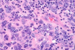
 Micrograph showing a follicular dendritic cell sarcoma. The (benign) interspersed lymphocytes are a characteristic finding. H&E stain. | |
|
| |
| Synonyms | follicular dendritic cell tumour, follicular dendritic cell neoplasm |
|
| |
| LM | oval or spindle-shaped cellular & nuclear morphology, variable architecture (sheets, fascicles, whorles, storiform pattern), nuclei with small nucleoli and clear or dispersed chromatin, multinucleated cells, interspersed small lymphocytes, +/-necrosis, +/-marked nuclear atypia, +/-abundant mitoses. |
| LM DDx | histiocytic sarcoma, thymoma, other spindle cell lesions |
| IHC | CD21 +ve, CD23 +ve, CD35 +ve, Ki-M4p +ve, Ki-FDRC1p +ve, vimentin +ve |
| Associated Dx | Castleman disease, hyaline-vascular type |
| Prevalence | very rare |
| Treatment | excision |
Follicular dendritic cell sarcoma, abbreviated FDCS, is a very rare malignant tumour.
It is also known as follicular dendritic cell tumour (abbreviated FDCT),[1] and follicular dendritic cell neoplasm.[2]
General
- Very rare.
- Behave like a low-grade sarcoma.[3]
- Reported association with Castleman disease (hyaline-vascular type).[3]
WHO 2001 classification of dendritic cell neoplasms:[4]
- Langerhans cell histiocytosis.
- Langerhans cell sarcoma.
- Interdigitating dendritic cell sarcoma/tumour.
- Follicular dendritic cell sarcoma/tumour.
- Dendritic cell sarcoma, not specified otherwise.
Gross
Features:
- Often a head and neck lesion.[citation needed]
Microscopic
Features:[3]
- Oval or spindle-shaped cellular & nuclear morphology.
- Variable architecture (sheets, fascicles, whorles, storiform pattern).
- Nuclei:
- Small nucleoli.
- Clear or dispersed chromatin.
- Multinucleated cells.
- Interspersed small lymphocytes - distinctive feature.
- +/-Necrosis.
- +/-Marked nuclear atypia.
- +/-Abundant mitoses.
DDx:
- Histiocytic sarcoma.[5]
- Thymoma.
- Spindle cell lesions such as sarcomatoid carcinoma.
- Meningioma.[citation needed]
Images
Related images
www
- FDCS (ajronline.org).[1]
- FDCS (npplweb.com).[6]
- FDCT - several crappy images (upmc.edu).
- FDCS - several images (upmc.edu).
IHC
Features:[3]
- CD21 +ve.
- CD35 +ve.
- Ki-M4p +ve
- Ki-FDRC1p +ve.
- Vimentin +ve.
- S-100 +ve/-ve.
- Muscle-specific actin +ve/-ve.
- EMA +ve/-ve.
Additional stains:[7]
- CD23 +ve.
- D2-40 +ve.
Note:
- The interspersed lymphocytes are B cells.[8]
Images
See also
References
- ↑ 1.0 1.1 Leipsic, JA.; McAdams, HP.; Sporn, TA. (Jun 2007). "Follicular dendritic cell sarcoma of the mediastinum.". AJR Am J Roentgenol 188 (6): W554-6. doi:10.2214/AJR.04.1530. PMID 17515347.
- ↑ Denning, KL.; Olson, PR.; Maley, RH.; Flati, VR.; Myers, JL.; Silverman, JF. (Apr 2009). "Primary pulmonary follicular dendritic cell neoplasm: a case report and review of the literature.". Arch Pathol Lab Med 133 (4): 643-7. doi:10.1043/1543-2165-133.4.643. PMID 19391666.
- ↑ 3.0 3.1 3.2 3.3 Perez-Ordoñez, B.; Rosai, J. (May 1998). "Follicular dendritic cell tumor: review of the entity.". Semin Diagn Pathol 15 (2): 144-54. PMID 9606805.
- ↑ Kairouz, S.; Hashash, J.; Kabbara, W.; McHayleh, W.; Tabbara, IA. (Oct 2007). "Dendritic cell neoplasms: an overview.". Am J Hematol 82 (10): 924-8. doi:10.1002/ajh.20857. PMID 17636477.
- ↑ Alexiev, BA.; Sailey, CJ.; McClure, SA.; Ord, RA.; Zhao, XF.; Papadimitriou, JC. (2007). "Primary histiocytic sarcoma arising in the head and neck with predominant spindle cell component.". Diagn Pathol 2: 7. doi:10.1186/1746-1596-2-7. PMID 17324277. http://www.diagnosticpathology.org/content/2/1/7.
- ↑ URL: http://www.npplweb.com/wjsmro/fulltext/2/1. Accessed on: September 13, 2014.
- ↑ Yin, WH.; Yu, GY.; Ma, Y.; Rao, HL.; Lin, SX.; Shao, CK.; Liang, Q.; Guo, N. et al. (Aug 2010). "[Follicular dendritic cell sarcoma: a clinicopathologic analysis of ten cases].". Zhonghua Bing Li Xue Za Zhi 39 (8): 522-7. PMID 21055030.
- ↑ Lorenzi, L.; Lonardi, S.; Petrilli, G.; Tanda, F.; Bella, M.; Laurino, L.; Rossi, G.; Facchetti, F. (Feb 2012). "Folliculocentric B-cell-rich follicular dendritic cells sarcoma: a hitherto unreported morphological variant mimicking lymphoproliferative disorders.". Hum Pathol 43 (2): 209-15. doi:10.1016/j.humpath.2011.02.029. PMID 21835430.


