Difference between revisions of "Asbestosis"
Jump to navigation
Jump to search
(→Images) |
|||
| (2 intermediate revisions by the same user not shown) | |||
| Line 16: | Line 16: | ||
| Grossing = | | Grossing = | ||
| Site = [[lung]] - see ''[[diffuse lung diseases]]'' | | Site = [[lung]] - see ''[[diffuse lung diseases]]'' | ||
| Assdx = [[pleural plaques]], pleural effusion (may be benign), [[malignant mesothelioma]] | | Assdx = [[pleural plaques]], [[pleural effusion]] (may be benign), [[malignant mesothelioma]] | ||
| Syndromes = | | Syndromes = | ||
| Clinicalhx = exposure to asbestos | | Clinicalhx = exposure to asbestos | ||
| Line 47: | Line 47: | ||
Diagnosis: | Diagnosis: | ||
*Rests on morphology with special techniques (e.g. polarization)<ref>{{Cite journal | last1 = Galateau-Salle | first1 = F. | title = [Anatomopathological tools for screening and medical surveillance of people exposed to asbestos]. | journal = Rev Mal Respir | volume = 16 | issue = 6 Pt 2 | pages = 1244-56 | month = Dec | year = 1999 | doi = | PMID = 10897845 }}</ref> and includes tunneling [[electron microscopy]] (TEM),<ref name=pmid21479897>{{Cite journal | last1 = Neumann | first1 = V. | last2 = Löseke | first2 = S. | last3 = Tannapfel | first3 = A. | title = Mesothelioma and analysis of tissue fiber content. | journal = Recent Results Cancer Res | volume = 189 | issue = | pages = 79-95 | month = | year = 2011 | doi = 10.1007/978-3-642-10862-4_6 | PMID = 21479897 }}</ref> as done at special centres. | *Rests on morphology with special techniques (e.g. [[polarization]])<ref name=pmid10897845>{{Cite journal | last1 = Galateau-Salle | first1 = F. | title = [Anatomopathological tools for screening and medical surveillance of people exposed to asbestos]. | journal = Rev Mal Respir | volume = 16 | issue = 6 Pt 2 | pages = 1244-56 | month = Dec | year = 1999 | doi = | PMID = 10897845 }}</ref> and includes tunneling [[electron microscopy]] (TEM),<ref name=pmid21479897>{{Cite journal | last1 = Neumann | first1 = V. | last2 = Löseke | first2 = S. | last3 = Tannapfel | first3 = A. | title = Mesothelioma and analysis of tissue fiber content. | journal = Recent Results Cancer Res | volume = 189 | issue = | pages = 79-95 | month = | year = 2011 | doi = 10.1007/978-3-642-10862-4_6 | PMID = 21479897 }}</ref> as done at special centres. | ||
**Tissue is typically digested prior to fibre counting.<ref name=pmid20196674>{{Cite journal | last1 = Roggli | first1 = VL. | last2 = Gibbs | first2 = AR. | last3 = Attanoos | first3 = R. | last4 = Churg | first4 = A. | last5 = Popper | first5 = H. | last6 = Cagle | first6 = P. | last7 = Corrin | first7 = B. | last8 = Franks | first8 = TJ. | last9 = Galateau-Salle | first9 = F. | title = Pathology of asbestosis- An update of the diagnostic criteria: Report of the asbestosis committee of the college of american pathologists and pulmonary pathology society. | journal = Arch Pathol Lab Med | volume = 134 | issue = 3 | pages = 462-80 | month = Mar | year = 2010 | doi = 10.1043/1543-2165-134.3.462 | PMID = 20196674 }}</ref> | **Tissue is typically digested prior to fibre counting.<ref name=pmid20196674>{{Cite journal | last1 = Roggli | first1 = VL. | last2 = Gibbs | first2 = AR. | last3 = Attanoos | first3 = R. | last4 = Churg | first4 = A. | last5 = Popper | first5 = H. | last6 = Cagle | first6 = P. | last7 = Corrin | first7 = B. | last8 = Franks | first8 = TJ. | last9 = Galateau-Salle | first9 = F. | title = Pathology of asbestosis- An update of the diagnostic criteria: Report of the asbestosis committee of the college of american pathologists and pulmonary pathology society. | journal = Arch Pathol Lab Med | volume = 134 | issue = 3 | pages = 462-80 | month = Mar | year = 2010 | doi = 10.1043/1543-2165-134.3.462 | PMID = 20196674 }}</ref> | ||
| Line 53: | Line 53: | ||
Features:<ref name=pmid20196674/> | Features:<ref name=pmid20196674/> | ||
#Interstitial fibrosis - similar to [[usual interstitial pneumonia]] (UIP). | #Interstitial fibrosis - similar to [[usual interstitial pneumonia]] (UIP). | ||
#''Ferruginous bodies'' - '''key feature'''. | #''[[Ferruginous bodies]]'' - '''key feature'''. | ||
#*Segmented twirling baton with long slender fibre within. | #*Segmented twirling baton with long slender fibre within. | ||
#*Black crystal-like appearance - may be confused with dirt on H&E. | #*Black crystal-like appearance - may be confused with dirt on H&E. | ||
Latest revision as of 20:15, 23 April 2016
| Asbestosis | |
|---|---|
| Diagnosis in short | |
 Asbestosis. H&E stain. | |
|
| |
| LM | interstitial fibrosis (similar to usual interstitial pneumonia), ferruginous bodies (segmented twirling baton with long slender fibre within it) |
| LM DDx | usual interstitial pneumonia |
| Stains | Iron stain +ve (ferruginous bodies) |
| Site | lung - see diffuse lung diseases |
|
| |
| Associated Dx | pleural plaques, pleural effusion (may be benign), malignant mesothelioma |
| Clinical history | exposure to asbestos |
| Prevalence | uncommon |
| Radiology | interstitial pattern, lower lobe predominant |
| Prognosis | poor |
Asbestosis is a diffuse lung disease due to asbestos exposure. It is a type of hypersensitivity pneumonitis.[1]
General
Definition:
- Interstitial lung disease due to asbestos exposure.[2]
- Important to diagnose... asbestosis = compensation.
Conditions associated with asbestos exposure (mnemonic PALM):[2]
- Pleural plaques.
- Asbestosis.
- Lung carcinoma.
- Malignant mesothelioma.
Possible association with asbestos exposure:
Diagnosis:
- Rests on morphology with special techniques (e.g. polarization)[4] and includes tunneling electron microscopy (TEM),[5] as done at special centres.
- Tissue is typically digested prior to fibre counting.[6]
Microscopic
Features:[6]
- Interstitial fibrosis - similar to usual interstitial pneumonia (UIP).
- Ferruginous bodies - key feature.
- Segmented twirling baton with long slender fibre within.
- Black crystal-like appearance - may be confused with dirt on H&E.
Images
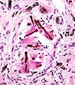
Ferruginous bodies. (WC)
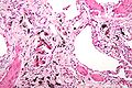
Asbestosis. (WC)
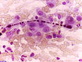
Ferruginous body in carcinoma - cytology. (WC/Alex Brollo)


Stains
- Iron stain +ve.
See also
References
- ↑ Zacharisen MC, Fink JN (November 2011). "Hypersensitivity pneumonitis and related conditions in the work environment". Immunol Allergy Clin North Am 31 (4): 769–86, vii. doi:10.1016/j.iac.2011.07.004. PMID 21978856.
- ↑ 2.0 2.1 Mitchell, Richard; Kumar, Vinay; Fausto, Nelson; Abbas, Abul K.; Aster, Jon (2011). Pocket Companion to Robbins & Cotran Pathologic Basis of Disease (8th ed.). Elsevier Saunders. pp. 375. ISBN 978-1416054542.
- ↑ Reid, A.; Heyworth, J.; de Klerk, N.; Musk, AW. (Nov 2009). "Asbestos exposure and gestational trophoblastic disease: a hypothesis.". Cancer Epidemiol Biomarkers Prev 18 (11): 2895-8. doi:10.1158/1055-9965.EPI-09-0731. PMID 19900938.
- ↑ Galateau-Salle, F. (Dec 1999). "[Anatomopathological tools for screening and medical surveillance of people exposed to asbestos].". Rev Mal Respir 16 (6 Pt 2): 1244-56. PMID 10897845.
- ↑ Neumann, V.; Löseke, S.; Tannapfel, A. (2011). "Mesothelioma and analysis of tissue fiber content.". Recent Results Cancer Res 189: 79-95. doi:10.1007/978-3-642-10862-4_6. PMID 21479897.
- ↑ 6.0 6.1 Roggli, VL.; Gibbs, AR.; Attanoos, R.; Churg, A.; Popper, H.; Cagle, P.; Corrin, B.; Franks, TJ. et al. (Mar 2010). "Pathology of asbestosis- An update of the diagnostic criteria: Report of the asbestosis committee of the college of american pathologists and pulmonary pathology society.". Arch Pathol Lab Med 134 (3): 462-80. doi:10.1043/1543-2165-134.3.462. PMID 20196674.