Difference between revisions of "Chiari malformation"
Jump to navigation
Jump to search
Jensflorian (talk | contribs) (histology in chiari (mostly type II)) |
|||
| (2 intermediate revisions by one other user not shown) | |||
| Line 4: | Line 4: | ||
*Usually a radiologic diagnosis. | *Usually a radiologic diagnosis. | ||
*May be seen in a [[fetal autopsy]]. | *May be seen in a [[fetal autopsy]]. | ||
Clinical:<ref>{{Cite journal | last1 = Ferré Masó | first1 = A. | last2 = Poca | first2 = MA. | last3 = de la Calzada | first3 = MD. | last4 = Solana | first4 = E. | last5 = Romero Tomás | first5 = O. | last6 = Sahuquillo | first6 = J. | title = Sleep disturbance: a forgotten syndrome in patients with Chiari I malformation. | journal = Neurologia | volume = | issue = | pages = | month = Mar | year = 2011 | doi = 10.1016/j.nrl.2011.01.008 | PMID = 21420201 }}</ref> | |||
*Headaches, occipital. | |||
*Dizziness. | |||
*Nocturnal respiratory abnormalities. | |||
===Classification=== | ===Classification=== | ||
Numbered from least severe to most severe: | Numbered from least severe to most severe: | ||
*Chiari type I - tonsils herniated<ref>URL: [http://rarediseases.info.nih.gov/GARD/Disease.aspx?diseaseID=9230 http://rarediseases.info.nih.gov/GARD/Disease.aspx?diseaseID=9230]. Accessed on: 6 May 2011.</ref> (radiologic definition: 4-6 mm below the plane of the foramen magnum). | *Chiari type I - tonsils herniated<ref>URL: [http://rarediseases.info.nih.gov/GARD/Disease.aspx?diseaseID=9230 http://rarediseases.info.nih.gov/GARD/Disease.aspx?diseaseID=9230]. Accessed on: 6 May 2011.</ref> (radiologic definition: 4-6 mm below the plane of the foramen magnum). | ||
**Associated with: sudden death, sleep apnea, cerebellar ataxia. | **Associated with: sudden death,<ref>{{Cite journal | last1 = Zhang | first1 = J. | last2 = Shao | first2 = Y. | last3 = Qin | first3 = Z. | last4 = Liu | first4 = N. | last5 = Zou | first5 = D. | last6 = Huang | first6 = P. | last7 = Chen | first7 = Y. | title = Sudden Unexpected Death due to Chiari Type I Malformation in a Road Accident Case. | journal = J Forensic Sci | volume = | issue = | pages = | month = Dec | year = 2012 | doi = 10.1111/1556-4029.12051 | PMID = 23278920 }}</ref> sleep apnea, cerebellar ataxia. | ||
*Chiari type II - often assoc. with hydrocephaly at birth. | *Chiari type II - often assoc. with hydrocephaly at birth. Often associated with [[myelomeningocele]]. | ||
*Chiari type III - cerebellum + brain stem herniate through foramen magnum +/- encephalocele.<ref>URL: [http://www.ninds.nih.gov/disorders/chiari/detail_chiari.htm http://www.ninds.nih.gov/disorders/chiari/detail_chiari.htm]. Accessed on: 6 May 2011.</ref> | *Chiari type III - cerebellum + brain stem herniate through foramen magnum +/- encephalocele.<ref>URL: [http://www.ninds.nih.gov/disorders/chiari/detail_chiari.htm http://www.ninds.nih.gov/disorders/chiari/detail_chiari.htm]. Accessed on: 6 May 2011.</ref> | ||
*Chiari type IV - cerebellar hypoplasia or no cerebellum. | *Chiari type IV - cerebellar hypoplasia or no cerebellum. | ||
==Histology== | |||
Surgery depends on clinical symptoms due to CSF obstructions. Specimens may include: <ref>{{Cite journal | last1 = Piatt | first1 = JH. | last2 = D'Agostino | first2 = A. | title = The Chiari II malformation: lesions discovered within the fourth ventricle. | journal = Pediatr Neurosurg | volume = 30 | issue = 2 | pages = 79-85 | month = Feb | year = 1999 | doi = 28767 | PMID = 10325563 }}</ref> | |||
* Vertebral bone (decompression) | |||
* Hermiated cerebellar tonsils. | |||
* Reactive / dysplastic [[choroid plexus]]. | |||
* Glial nodules. | |||
* Arachnoidal cysts. | |||
* [[Subependymoma]]s. | |||
In cases with [[myelomeningocele]] at autopsy, the posterior fossa should be examined. | |||
==Images== | |||
<gallery> | |||
File:Chiari2.jpg|Schematic display of Chiari type II malformation. | |||
File:Chiari-Malformation_MRT_T2_sag.jpg | Radiology of Chiari type II. | |||
File:Hypertrophic_plexus_chiari_II_low_mag.jpg | Hypertrophic plexus choroideus in Chiari type II (low mag). | |||
File:Hypertrophic_plexus_chiari_II_intermed_mag.jpg | Fibrous tissue within a hypertrophic plexus choroideus in Chiari type II (intermed mag). | |||
</gallery> | |||
==See also== | ==See also== | ||
| Line 20: | Line 43: | ||
{{Reflist|2}} | {{Reflist|2}} | ||
[[Category:Diagnosis]] | |||
[[Category:Neuropathology]] | [[Category:Neuropathology]] | ||
Latest revision as of 09:12, 16 September 2015
Chiari malformation is a developmental abnormality of the brain.
General
- Usually a radiologic diagnosis.
- May be seen in a fetal autopsy.
Clinical:[1]
- Headaches, occipital.
- Dizziness.
- Nocturnal respiratory abnormalities.
Classification
Numbered from least severe to most severe:
- Chiari type I - tonsils herniated[2] (radiologic definition: 4-6 mm below the plane of the foramen magnum).
- Associated with: sudden death,[3] sleep apnea, cerebellar ataxia.
- Chiari type II - often assoc. with hydrocephaly at birth. Often associated with myelomeningocele.
- Chiari type III - cerebellum + brain stem herniate through foramen magnum +/- encephalocele.[4]
- Chiari type IV - cerebellar hypoplasia or no cerebellum.
Histology
Surgery depends on clinical symptoms due to CSF obstructions. Specimens may include: [5]
- Vertebral bone (decompression)
- Hermiated cerebellar tonsils.
- Reactive / dysplastic choroid plexus.
- Glial nodules.
- Arachnoidal cysts.
- Subependymomas.
In cases with myelomeningocele at autopsy, the posterior fossa should be examined.
Images
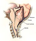
Schematic display of Chiari type II malformation.

Radiology of Chiari type II.

Hypertrophic plexus choroideus in Chiari type II (low mag).

Fibrous tissue within a hypertrophic plexus choroideus in Chiari type II (intermed mag).


See also
References
- ↑ Ferré Masó, A.; Poca, MA.; de la Calzada, MD.; Solana, E.; Romero Tomás, O.; Sahuquillo, J. (Mar 2011). "Sleep disturbance: a forgotten syndrome in patients with Chiari I malformation.". Neurologia. doi:10.1016/j.nrl.2011.01.008. PMID 21420201.
- ↑ URL: http://rarediseases.info.nih.gov/GARD/Disease.aspx?diseaseID=9230. Accessed on: 6 May 2011.
- ↑ Zhang, J.; Shao, Y.; Qin, Z.; Liu, N.; Zou, D.; Huang, P.; Chen, Y. (Dec 2012). "Sudden Unexpected Death due to Chiari Type I Malformation in a Road Accident Case.". J Forensic Sci. doi:10.1111/1556-4029.12051. PMID 23278920.
- ↑ URL: http://www.ninds.nih.gov/disorders/chiari/detail_chiari.htm. Accessed on: 6 May 2011.
- ↑ Piatt, JH.; D'Agostino, A. (Feb 1999). "The Chiari II malformation: lesions discovered within the fourth ventricle.". Pediatr Neurosurg 30 (2): 79-85. doi:28767. PMID 10325563.