Sjögren syndrome: Difference between revisions
small fix, note redirect |
|||
| (15 intermediate revisions by the same user not shown) | |||
| Line 2: | Line 2: | ||
The syndrome may be part of another [[connective tissue disorder]], e.g. [[rheumatoid arthritis]], in which case it is called ''secondary Sjögren syndrome''.<ref name=pmid1703737>{{Cite journal | last1 = Celenligil | first1 = H. | last2 = Kansu | first2 = E. | last3 = Ruacan | first3 = S. | last4 = Eratalay | first4 = K. | last5 = Irkeç | first5 = M. | title = Characterization of peripheral blood and salivary gland lymphocytes in secondary Sjögren's syndrome. | journal = Ann Dent | volume = 49 | issue = 2 | pages = 18-22 | month = | year = 1990 | doi = | PMID = 1703737 }}</ref> | The syndrome may be part of another [[connective tissue disorder]], e.g. [[rheumatoid arthritis]], in which case it is called ''secondary Sjögren syndrome''.<ref name=pmid1703737>{{Cite journal | last1 = Celenligil | first1 = H. | last2 = Kansu | first2 = E. | last3 = Ruacan | first3 = S. | last4 = Eratalay | first4 = K. | last5 = Irkeç | first5 = M. | title = Characterization of peripheral blood and salivary gland lymphocytes in secondary Sjögren's syndrome. | journal = Ann Dent | volume = 49 | issue = 2 | pages = 18-22 | month = | year = 1990 | doi = | PMID = 1703737 }}</ref> | ||
Pathologists are likely to see this condition as a ''[[labial salivary gland]]'' biopsy. | |||
''Lip biopsy'' redirects here. | |||
==General== | ==General== | ||
| Line 51: | Line 55: | ||
Features ([[salivary gland]]):<ref name=pmid19323360/> | Features ([[salivary gland]]):<ref name=pmid19323360/> | ||
*"Significant lymphocytic infiltrate" adjacent to viable [[salivary gland]] (or [[lacrimal gland]]) acini - '''key feature'''. | *"Significant lymphocytic infiltrate" adjacent to viable [[salivary gland]] (or [[lacrimal gland]]) acini - '''key feature'''. | ||
**"Significant | **"Significant lymphocytic infiltrate": cluster of >= 50 lymphocytes - '''important'''. | ||
***Lymphocytes may be perivascular or periductular.<ref name=pmid12022353/><ref name=pmid8003059>{{Cite journal | last1 = Daniels | first1 = TE. | last2 = Whitcher | first2 = JP. | title = Association of patterns of labial salivary gland inflammation with keratoconjunctivitis sicca. Analysis of 618 patients with suspected Sjögren's syndrome. | journal = Arthritis Rheum | volume = 37 | issue = 6 | pages = 869-77 | month = Jun | year = 1994 | doi = | PMID = 8003059 }}</ref> | ***Lymphocytes may be perivascular or periductular.<ref name=pmid12022353/><ref name=pmid8003059>{{Cite journal | last1 = Daniels | first1 = TE. | last2 = Whitcher | first2 = JP. | title = Association of patterns of labial salivary gland inflammation with keratoconjunctivitis sicca. Analysis of 618 patients with suspected Sjögren's syndrome. | journal = Arthritis Rheum | volume = 37 | issue = 6 | pages = 869-77 | month = Jun | year = 1994 | doi = | PMID = 8003059 }}</ref> | ||
***[[Plasma cell]]s should not exceed 10% of the inflammatory infiltrate.<ref name=pmid12022353/> | ***[[Plasma cell]]s should not exceed 10% of the inflammatory infiltrate.<ref name=pmid12022353/> | ||
| Line 64: | Line 68: | ||
*Diagnosis is based on clinicopathologic correlation; the histology alone is insufficient. | *Diagnosis is based on clinicopathologic correlation; the histology alone is insufficient. | ||
Images: | ===Images=== | ||
<gallery> | |||
Image:Sjogren_syndrome_%281%29.jpg | SS - low mag. (WC) | |||
Image:Sjogren_syndrome_%282%29.jpg | SS - high mag. (WC) | |||
</gallery> | |||
www: | |||
*[http://img.medscape.com/pi/emed/ckb/rheumatology/329097-1339496-332125-1582482.jpg Sjögren syndrome (medscape.com)].<ref>URL: [http://emedicine.medscape.com/article/332125-workup#aw2aab6b5b6aa http://emedicine.medscape.com/article/332125-workup#aw2aab6b5b6aa]. Accessed on: 24 July 2012.</ref> | *[http://img.medscape.com/pi/emed/ckb/rheumatology/329097-1339496-332125-1582482.jpg Sjögren syndrome (medscape.com)].<ref>URL: [http://emedicine.medscape.com/article/332125-workup#aw2aab6b5b6aa http://emedicine.medscape.com/article/332125-workup#aw2aab6b5b6aa]. Accessed on: 24 July 2012.</ref> | ||
===Focus score=== | ===Focus score=== | ||
Features:<ref name=pmid12022353/> | Features:<ref name=pmid12022353/> | ||
*This is nothing more than a count of significant lymphocytic foci. | *This is nothing more than a count of significant lymphocytic foci per 4 mm*mm. | ||
**Significant: >= 50 lymphocytes, adjacent to salivary gland acini. | **Significant: >= 50 lymphocytes, adjacent to (viable) [[salivary gland]] acini. | ||
** | **A ''focus score'' of one or more is considered significant.<ref name=pmid12006334/> | ||
Calculating the focus score (fs): | |||
# Count the significant foci (n). | |||
# Estimate the sample area in mm<sup>2</sup> (a). | |||
:<math>fs = { n \over a } \times 4 \ mm^2</math> | |||
====Grading==== | ====Grading==== | ||
| Line 103: | Line 116: | ||
==Sign out== | ==Sign out== | ||
===Suggestive=== | |||
<pre> | <pre> | ||
LOWER LIP, BIOPSY: | LOWER LIP, BIOPSY: | ||
- | - MINOR SALIVARY GLAND WITH FOCAL LYMPHOCYTIC SIALADENITIS, SEE COMMENT. | ||
- SQUAMOUS MUCOSA WITH PARAKERATOSIS, MILD. | |||
- | |||
COMMENT: | COMMENT: | ||
The | The histologic findings are compatible with those seen in Sjoegren's disease | ||
(focus score >= 1). | |||
is required. | |||
SALIVARY GLAND - SUMMARY: | |||
Glandular area: 8 mm*mm. | |||
Interstitial fat: not apparent. | |||
Plasma cells: not apparent. | |||
Fibrosis: none apparent (0% of salivary gland area). | |||
Ductular dilation: not apparent. | |||
Gross foci: 4. | |||
Focus score (foci/4 mm*mm): 2. | |||
This result needs to be combined with the clinical and serologic criteria used | |||
to diagnose Sjoegren's disease. | |||
</pre> | |||
===Not suggestive=== | |||
<pre> | |||
Minor Salivary Gland, Biopsy: | |||
- Benign minor salivary gland with one focus of chronic lymphocyte | |||
predominant inflammation. | |||
- Focus score less than one; not suggestive of Sjoegren's disease. | |||
Comment: | |||
Estimated area of salivary gland: 50 mm*mm. | |||
Number of foci (~50 lymphocytes): 1. | |||
Plasma cells: few (<10%). | |||
Fibrosis: none/minimal. | |||
Focus score (number of foci / area x 4 mm*mm) = < 1. | |||
The findings do not exclude Sjoegren's disease. Clinical and serologic | |||
correlation is required. | |||
</pre> | </pre> | ||
Latest revision as of 21:45, 2 March 2023
Sjögren syndrome, also Sjögren disease, is an uncommon disease that rheumatologists see. Sjögren is also spelled Sjoegren and Sjogren.
The syndrome may be part of another connective tissue disorder, e.g. rheumatoid arthritis, in which case it is called secondary Sjögren syndrome.[1]
Pathologists are likely to see this condition as a labial salivary gland biopsy.
Lip biopsy redirects here.
General
Clinical - classically:
- Women in 50s.
- Dry mouth (xerostomia).
- Dry eyes (xerophthalmia).
Diagnostic criteria
European criteria of 2002:[2]
| Criteria | Details | Type |
|---|---|---|
| Oral symptoms | any: (1) dry mouth > 3 months, (2) require fluids for swallowing, (3) swollen salivary glands (adults) | history |
| Oral signs | any: (1) low salivary flow test positive, (2) salivary scintigraphy positive (3) (parotid) sialography positive | clinical test |
| Ocular symptoms | any: (1) dry eye > 3 months, (2) need artifical tears >3x/day, (3) sand or gravel in the eyes sensation | history |
| Ocular signs | any: (1) Schirmer's test positive, (2) ocular dye test positive | clinical test |
| Autoantibodies | anti-SSA/Ro and/or anti-SSB/La | serology |
| Histology | labial minor salivary gland biopsy focus score >= 1.0/ 4 mm*mm; definition: multiple lymphocytic foci with >50 lymphocytes adjacent to mucinous acini, evaluated in 4 mm*mm of glandular tissue | pathology |
The diagnosis is made if either:[2]
- Four of six criteria required, must include either autoantibodies or histology.
- Three of the four objective (non-history) criteria are met.
Notes:
- ANA[3] and RF[4] were criteria in the past; they are no longer considered important in the diagnosis.
Microscopic
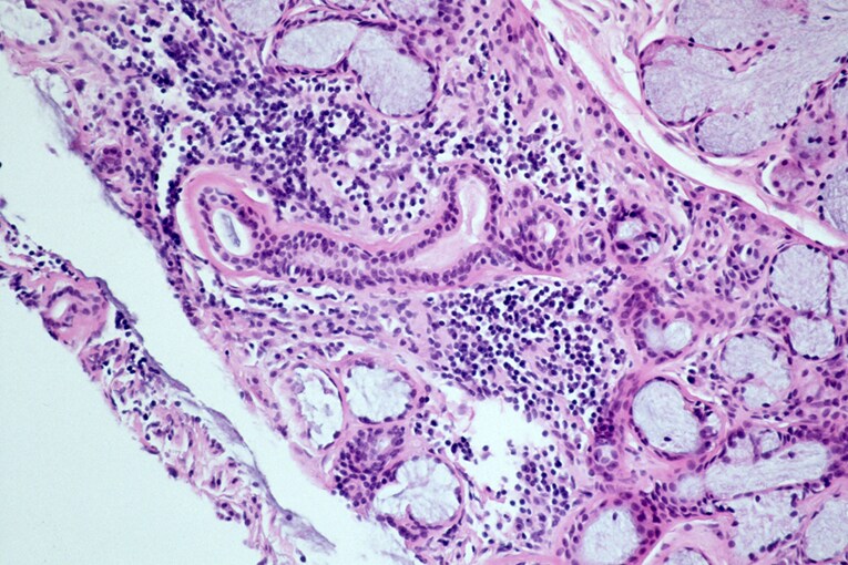
Features (salivary gland):[3]
- "Significant lymphocytic infiltrate" adjacent to viable salivary gland (or lacrimal gland) acini - key feature.
- "Significant lymphocytic infiltrate": cluster of >= 50 lymphocytes - important.
- Lymphocytes may be perivascular or periductular.[4][5]
- Plasma cells should not exceed 10% of the inflammatory infiltrate.[4]
- May have "benign lymphoepithelial lesions"[6] - intraepithelial lymphocytes.
- "Significant lymphocytic infiltrate": cluster of >= 50 lymphocytes - important.
- +/-Fibrosis.
DDx:
- MALT lymphoma.
- Chronic sialadenitis - mixed inflammatory infiltrate.
Note:
- Diagnosis is based on clinicopathologic correlation; the histology alone is insufficient.
Images
-
 SS - low mag. (WC)
 -
 SS - high mag. (WC)

.jpg)
www:
Focus score
Features:[4]
- This is nothing more than a count of significant lymphocytic foci per 4 mm*mm.
- Significant: >= 50 lymphocytes, adjacent to (viable) salivary gland acini.
- A focus score of one or more is considered significant.[2]
Calculating the focus score (fs):
- Count the significant foci (n).
- Estimate the sample area in mm2 (a).
Grading
In the past lesions were graded with the Chisholm-Mason classification.[6][8] The Chisholm-Mason classification is based on assessing 4 mm2 area of salivary gland tissue and depends on the abundance and aggregation of lymphocytes.
It is compares to Daniels-Whitcher grading as follows:
| Lymphocytes | Daniels-Whitcher grade |
Chisholm-Mason grade |
|---|---|---|
| slight infiltrate | mild | 1 |
| moderate infiltrate or less than one focus † | intermediate | 2 |
| one focus † | severe | 3 |
| more than one focus † | severe | 4 |
† Focus = an aggregrate of 50 lymphocytes or more.
Sign out
Suggestive
LOWER LIP, BIOPSY:
- MINOR SALIVARY GLAND WITH FOCAL LYMPHOCYTIC SIALADENITIS, SEE COMMENT.
- SQUAMOUS MUCOSA WITH PARAKERATOSIS, MILD.

COMMENT:
The histologic findings are compatible with those seen in Sjoegren's disease
(focus score >= 1).

SALIVARY GLAND - SUMMARY:
Glandular area: 8 mm*mm.
Interstitial fat: not apparent.
Plasma cells: not apparent.
Fibrosis: none apparent (0% of salivary gland area).
Ductular dilation: not apparent.
Gross foci: 4.
Focus score (foci/4 mm*mm): 2.

This result needs to be combined with the clinical and serologic criteria used
to diagnose Sjoegren's disease.
Not suggestive
Minor Salivary Gland, Biopsy:
- Benign minor salivary gland with one focus of chronic lymphocyte
  predominant inflammation.
- Focus score less than one; not suggestive of Sjoegren's disease.

Comment:
Estimated area of salivary gland: 50 mm*mm.
Number of foci (~50 lymphocytes): 1.
Plasma cells: few (<10%).
Fibrosis: none/minimal.
Focus score (number of foci / area x 4 mm*mm) = < 1.

The findings do not exclude Sjoegren's disease. Clinical and serologic
correlation is required.
See also
References
- ↑ Celenligil, H.; Kansu, E.; Ruacan, S.; Eratalay, K.; Irkeç, M. (1990). "Characterization of peripheral blood and salivary gland lymphocytes in secondary Sjögren's syndrome.". Ann Dent 49 (2): 18-22. PMID 1703737.
- ↑ 2.0 2.1 2.2 Vitali, C.; Bombardieri, S.; Jonsson, R.; Moutsopoulos, HM.; Alexander, EL.; Carsons, SE.; Daniels, TE.; Fox, PC. et al. (Jun 2002). "Classification criteria for Sjögren's syndrome: a revised version of the European criteria proposed by the American-European Consensus Group.". Ann Rheum Dis 61 (6): 554-8. PMC 1754137. PMID 12006334. https://www.ncbi.nlm.nih.gov/pmc/articles/PMC1754137/.
- ↑ 3.0 3.1 "Information from your family doctor. Sjögren syndrome.". Am Fam Physician 79 (6): 472. Mar 2009. PMID 19323360.
- ↑ 4.0 4.1 4.2 4.3 Vivino, FB.; Gala, I.; Hermann, GA. (May 2002). "Change in final diagnosis on second evaluation of labial minor salivary gland biopsies.". J Rheumatol 29 (5): 938-44. PMID 12022353.
- ↑ Daniels, TE.; Whitcher, JP. (Jun 1994). "Association of patterns of labial salivary gland inflammation with keratoconjunctivitis sicca. Analysis of 618 patients with suspected Sjögren's syndrome.". Arthritis Rheum 37 (6): 869-77. PMID 8003059.
- ↑ 6.0 6.1 Ramos-Casals, M.; Font, J. (Nov 2005). "Primary Sjögren's syndrome: current and emergent aetiopathogenic concepts.". Rheumatology (Oxford) 44 (11): 1354-67. doi:10.1093/rheumatology/keh714. PMID 15956090. http://rheumatology.oxfordjournals.org/content/44/11/1354.long.
- ↑ URL: http://emedicine.medscape.com/article/332125-workup#aw2aab6b5b6aa. Accessed on: 24 July 2012.
- ↑ Chisholm, DM.; Mason, DK. (Sep 1968). "Labial salivary gland biopsy in Sjögren's disease.". J Clin Pathol 21 (5): 656-60. PMC 473887. PMID 5697370. http://www.ncbi.nlm.nih.gov/pmc/articles/PMC473887/?tool=pubmed.