Difference between revisions of "Urothelial carcinoma in situ"
(→IHC) |
(→Images) |
||
| (5 intermediate revisions by the same user not shown) | |||
| Line 1: | Line 1: | ||
{{ Infobox diagnosis | {{ Infobox diagnosis | ||
| Name = {{PAGENAME}} | | Name = {{PAGENAME}} | ||
| Image = Urothelial carcinoma in situ -- very high mag.jpg | | Image = Urothelial carcinoma in situ -- very high mag.jpg | ||
| Width = | | Width = | ||
| Caption = Urothelial carcinoma in situ. [[H&E stain]]. | | Caption = Urothelial carcinoma in situ. [[H&E stain]]. | ||
| Line 24: | Line 24: | ||
| Bloodwork = | | Bloodwork = | ||
| Rads = | | Rads = | ||
| Endoscopy = | | Endoscopy = erythema or edema, may be unremarkable | ||
| Prognosis = | | Prognosis = | ||
| Other = | | Other = | ||
| ClinDDx = invasive (flat) [[urothelial carcinoma]] | | ClinDDx = invasive (flat) [[urothelial carcinoma]], inflammation (cystitis) | ||
| Tx = | | Tx = | ||
}} | }} | ||
| Line 47: | Line 47: | ||
*Urothelial carcinoma in situ (high-grade dysplasia). | *Urothelial carcinoma in situ (high-grade dysplasia). | ||
*Invasive [[urothelial carcinoma]]. | *Invasive [[urothelial carcinoma]]. | ||
==Gross== | |||
*Flat lesion - erythema or edema, may be unremarkable.<ref name=pmid19176205>{{Cite journal | last1 = Nese | first1 = N. | last2 = Gupta | first2 = R. | last3 = Bui | first3 = MH. | last4 = Amin | first4 = MB. | title = Carcinoma in situ of the urinary bladder: review of clinicopathologic characteristics with an emphasis on aspects related to molecular diagnostic techniques and prognosis. | journal = J Natl Compr Canc Netw | volume = 7 | issue = 1 | pages = 48-57 | month = Jan | year = 2009 | doi = | PMID = 19176205 }}</ref> | |||
==Microscopic== | ==Microscopic== | ||
| Line 70: | Line 73: | ||
===Images=== | ===Images=== | ||
<gallery> | <gallery> | ||
Image: Urothelial carcinoma in situ -- intermed mag.jpg | UCIS - intermed. mag. | Image: Urothelial carcinoma in situ -- intermed mag.jpg | UCIS - intermed. mag. (WC) | ||
Image: Urothelial carcinoma in situ -- high mag.jpg | UCIS - high mag. | Image: Urothelial carcinoma in situ -- high mag.jpg | UCIS - high mag. (WC) | ||
Image: Urothelial carcinoma in situ - alt -- high mag.jpg | UCIS - high mag. | Image: Urothelial carcinoma in situ - alt -- high mag.jpg | UCIS - high mag. (WC) | ||
Image: Urothelial carcinoma in situ -- very high mag.jpg | UCIS - very high mag. | Image: Urothelial carcinoma in situ -- very high mag.jpg | UCIS - very high mag. (WC) | ||
</gallery> | </gallery> | ||
www | <gallery> | ||
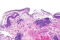
Image: Urothelial CIS -- low mag.jpg | UCIS - low mag. (WC) | |||
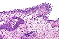
Image: Urothelial CIS -- intermed mag.jpg | UCIS - intermed. mag. (WC) | |||
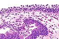
Image: Urothelial CIS -- high mag.jpg | UCIS - high mag. (WC) | |||
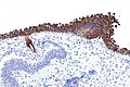
Image: Urothelial CIS - CK20 -- intermed mag.jpg | UCIS - CK20 - intermed. mag. (WC) | |||
Image: Urothelial CIS - p63 -- intermed mag.jpg | UCIS - p63 - intermed. mag. (WC) | |||
Image: Urothelial CIS - Ki67 -- high mag.jpg | UCIS - Ki-67 - high mag. (WC) | |||
</gallery> | |||
====www==== | |||
*[http://www.webpathology.com/image.asp?n=7&Case=57 Urothelial CIS (webpathology.com)]. | *[http://www.webpathology.com/image.asp?n=7&Case=57 Urothelial CIS (webpathology.com)]. | ||
*[http://www.webpathology.com/image.asp?n=10&Case=57 Urothelial CIS with shedding (webpathology.com)]. | *[http://www.webpathology.com/image.asp?n=10&Case=57 Urothelial CIS with shedding (webpathology.com)]. | ||
| Line 96: | Line 108: | ||
*CD44 -ve.<ref name=pmid24225842>{{Cite journal | last1 = Aron | first1 = M. | last2 = Luthringer | first2 = DJ. | last3 = McKenney | first3 = JK. | last4 = Hansel | first4 = DE. | last5 = Westfall | first5 = DE. | last6 = Parakh | first6 = R. | last7 = Mohanty | first7 = SK. | last8 = Balzer | first8 = B. | last9 = Amin | first9 = MB. | title = Utility of a triple antibody cocktail intraurothelial neoplasm-3 (IUN-3-CK20/CD44s/p53) and α-methylacyl-CoA racemase (AMACR) in the distinction of urothelial carcinoma in situ (CIS) and reactive urothelial atypia. | journal = Am J Surg Pathol | volume = 37 | issue = 12 | pages = 1815-23 | month = Dec | year = 2013 | doi = 10.1097/PAS.0000000000000114 | PMID = 24225842 }}</ref> | *CD44 -ve.<ref name=pmid24225842>{{Cite journal | last1 = Aron | first1 = M. | last2 = Luthringer | first2 = DJ. | last3 = McKenney | first3 = JK. | last4 = Hansel | first4 = DE. | last5 = Westfall | first5 = DE. | last6 = Parakh | first6 = R. | last7 = Mohanty | first7 = SK. | last8 = Balzer | first8 = B. | last9 = Amin | first9 = MB. | title = Utility of a triple antibody cocktail intraurothelial neoplasm-3 (IUN-3-CK20/CD44s/p53) and α-methylacyl-CoA racemase (AMACR) in the distinction of urothelial carcinoma in situ (CIS) and reactive urothelial atypia. | journal = Am J Surg Pathol | volume = 37 | issue = 12 | pages = 1815-23 | month = Dec | year = 2013 | doi = 10.1097/PAS.0000000000000114 | PMID = 24225842 }}</ref> | ||
**Positive in ''indeterminant'' and ''negative''. | **Positive in ''indeterminant'' and ''negative''. | ||
====Others==== | |||
*AMACR +ve (80% and 50% of untreated and treated CIS respectively<ref name=pmid24225842/>). | |||
===Images=== | ===Images=== | ||
Latest revision as of 04:56, 23 November 2016
Urothelial carcinoma in situ, also known as high-grade (urothelial) dysplasia, a non-invasive urothelial neoplasm without papillae.
| Urothelial carcinoma in situ | |
|---|---|
| Diagnosis in short | |
 Urothelial carcinoma in situ. H&E stain. | |
|
| |
| Synonyms | urothelial cell carcinoma in situ, high-grade dysplasia |
|
| |
| LM | nuclear changes (enlargement of nuclei (often 4-5x the size of stromal lymphocytes), nuclear pleomorphism - marked variation in size of nuclei), +/-disordered arrangement/crowding of cells, +/-mitoses, +/-enlarged nucleoli |
| LM DDx | urothelial carcinoma, urothelial dysplasia, urothelial atypia of unknown significance |
| IHC | CK20 +ve (full thickness), Ki-67 high, p53 +ve, CD44 -ve, CK7 +ve |
| Site | urothelium - urinary bladder, ureter, renal pelvis, prostatic urethra |
|
| |
| Signs | +/-hematuria |
| Prevalence | relatively uncommon |
| Endoscopy | erythema or edema, may be unremarkable |
| Clin. DDx | invasive (flat) urothelial carcinoma, inflammation (cystitis) |
It is also known as carcinoma in situ (abbreviated CIS) and urothelial cell carcinoma in situ (abbreviated UCC in situ). Urothelial carcinoma in situ may be abbreviated UCIS.
General
- Lack papillae.
- Uncommon in relation to other urothelial lesions.
- Less common than invasive flat urothelial carcinoma ~3-4x more common than UCIS.[1]
Classification of flat urothelial lesions
The World Health Organization classification is:[2]
- Reactive urothelial atypia.
- Flat urothelial hyperplasia.
- Urothelial atypia of unknown significance.
- Urothelial dysplasia (low-grade dysplasia).
- Urothelial carcinoma in situ (high-grade dysplasia).
- Invasive urothelial carcinoma.
Gross
- Flat lesion - erythema or edema, may be unremarkable.[3]
Microscopic
Features:
- Nuclear changes key feature.
- Enlargement of nuclei (often 4-5x the size of stromal lymphocytes) -- diagnostic.[4]
- Normal urothelium approx. 2x the size of stromal lymphocytes.
- Nuclear pleomorphism - marked variation in size of nuclei.
- Enlargement of nuclei (often 4-5x the size of stromal lymphocytes) -- diagnostic.[4]
- +/-Disordered arrangement/crowding of cells.
- In normal urothelium the cell line-up on the basement membrane.
- Umbrella cells often absent.
- +/-Mitoses present.
- +/-Enlarged nucleoli.
Note:
- The urothelium may be "depleted", i.e. exist only of rare large cells on the basement membrane.
- This is known as clinging urothelial carcinoma in situ.[5]
DDx:
- Urothelial atypia of unknown significance - waffle diagnosis.
- Urothelial dysplasia.
- Urothelial carcinoma, invasive.
Images
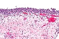
UCIS - intermed. mag. (WC)
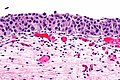
UCIS - high mag. (WC)
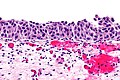
UCIS - high mag. (WC)
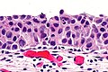
UCIS - very high mag. (WC)


UCIS - low mag. (WC)
UCIS - intermed. mag. (WC)
UCIS - high mag. (WC)
UCIS - CK20 - intermed. mag. (WC)
UCIS - p63 - intermed. mag. (WC)
UCIS - Ki-67 - high mag. (WC)


www
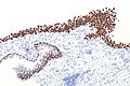
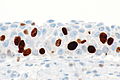
IHC
- CK20 +ve in deep cells.
- Normal urothelium -- only the umbrella cells.
- p53 +ve.
- CD44 -ve.
- Positive in indeterminant and negative.
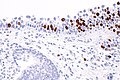
Another panel
Another panel for benign urothelium versus CIS:[7]
- CK20 +ve in deep cells (23/26 cases).
- Normal urothelium -- only the umbrella cells.
- Ki-67 ~50% of cells - deep and superficial.
- Normal ~10% of cells, confined to basal aspect.
- CD44 -ve.[8]
- Positive in indeterminant and negative.
Others
- AMACR +ve (80% and 50% of untreated and treated CIS respectively[8]).
Images
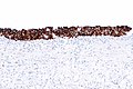
UCIS - CK20 - intermed. mag.
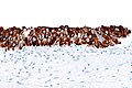
UCIS - CK20 - high mag.
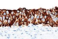
UCIS - CK20 - very high mag.
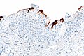
Benign urothelium - CK20 - high mag.


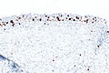
UCIS - Ki-67 - intermed. mag.
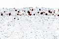
UCIS - Ki-67 - high mag.
UCIS - Ki-67 - very high mag.


Sign out
URINARY BLADDER LESION ("TUMOUR"), TRANSURETHRAL RESECTION URINARY BLADDER TUMOUR (TURBT):
- UROTHELIAL CARCINOMA IN SITU.
- BENIGN MUSCULARIS PROPRIA PRESENT.
URINARY BLADDER, RANDOM BIOPSIES:
- UROTHELIAL CARCINOMA IN SITU, SEE COMMENT.
-- NO EVIDENCE OF LAMINA PROPRIA INVASION.
- CHRONIC INFLAMMATION, MILD.
- BENIGN MUSCULARIS PROPRIA PRESENT.

COMMENT:
A CK20 immunostain marks the full thickness of the urothelium in atypical areas. A p53
immunostain moderately marks up to 20% of atypical cells focally.  A Ki-67 immunostain
marks 20-50% of the cells in the atypical areas.
Micro
The sections show multiple fragments of urothelium with nuclear hyperchromasia, nuclear crowding, mild-to-moderate nuclear enlargement, several atypical mitoses, and lack of maturation to the surface. There is no evidence of invasion. Benign muscularis propria is present.
See also
References
- ↑ Nielsen, ME.; Smith, AB.; Meyer, AM.; Kuo, TM.; Tyree, S.; Kim, WY.; Milowsky, MI.; Pruthi, RS. et al. (Jan 2014). "Trends in stage-specific incidence rates for urothelial carcinoma of the bladder in the United States: 1988 to 2006.". Cancer 120 (1): 86-95. doi:10.1002/cncr.28397. PMID 24122346.
- ↑ Hodges, KB.; Lopez-Beltran, A.; Davidson, DD.; Montironi, R.; Cheng, L. (Feb 2010). "Urothelial dysplasia and other flat lesions of the urinary bladder: clinicopathologic and molecular features.". Hum Pathol 41 (2): 155-62. doi:10.1016/j.humpath.2009.07.002. PMID 19762067.
- ↑ Nese, N.; Gupta, R.; Bui, MH.; Amin, MB. (Jan 2009). "Carcinoma in situ of the urinary bladder: review of clinicopathologic characteristics with an emphasis on aspects related to molecular diagnostic techniques and prognosis.". J Natl Compr Canc Netw 7 (1): 48-57. PMID 19176205.
- ↑ Zhou, Ming; Magi-Galluzzi, Cristina (2006). Genitourinary Pathology: A Volume in Foundations in Diagnostic Pathology Series (1st ed.). Churchill Livingstone. pp. 161. ISBN 978-0443066771.
- ↑ Amin, Mahul B. (2010). Diagnostic Pathology: Genitourinary (1st ed.). Amirsys. pp. 2-55. ISBN 978-1931884280.
- ↑ Amin MB, Epstein JI, Ulbright TM, et al. (August 2014). "Best practices recommendations in the application of immunohistochemistry in urologic pathology: report from the international society of urological pathology consensus conference". Am. J. Surg. Pathol. 38 (8): 1017–22. doi:10.1097/PAS.0000000000000254. PMID 25025364.
- ↑ Yin, H.; He, Q.; Li, T.; Leong, AS. (Sep 2006). "Cytokeratin 20 and Ki-67 to distinguish carcinoma in situ from flat non-neoplastic urothelium.". Appl Immunohistochem Mol Morphol 14 (3): 260-5. PMID 16932015.
- ↑ 8.0 8.1 Aron, M.; Luthringer, DJ.; McKenney, JK.; Hansel, DE.; Westfall, DE.; Parakh, R.; Mohanty, SK.; Balzer, B. et al. (Dec 2013). "Utility of a triple antibody cocktail intraurothelial neoplasm-3 (IUN-3-CK20/CD44s/p53) and α-methylacyl-CoA racemase (AMACR) in the distinction of urothelial carcinoma in situ (CIS) and reactive urothelial atypia.". Am J Surg Pathol 37 (12): 1815-23. doi:10.1097/PAS.0000000000000114. PMID 24225842.