Difference between revisions of "Helicobacter gastritis"
(create) |
(redirect) |
||
| (33 intermediate revisions by the same user not shown) | |||
| Line 1: | Line 1: | ||
# | {{ Infobox diagnosis | ||
| Name = {{PAGENAME}} | |||
| Image = Helicobacter gastritis - crop -- extremely high mag.jpg | |||
| Width = | |||
| Caption = Helicobacter gastritis. [[H&E stain]]. | |||
| Micro = helicobacter organisms, moderate chronic active gastritis (neutrophils esp. at the luminal aspect/intraepithelial, numerous plasma cell clusters) | |||
| Subtypes = ''[[Helicobacter pylori]]'', ''Helicobacter heilmannii'' | |||
| LMDDx = [[acute gastritis]], [[chronic gastritis]] | |||
| Stains = [[Diff-Quik]], [[Cresyl violet stain]], [[Warthin-Starry stain]] | |||
| IHC = Helicobacter IHC | |||
| EM = | |||
| Molecular = | |||
| IF = | |||
| Gross = | |||
| Grossing = | |||
| Site = [[stomach]] | |||
| Assdx = [[MALT lymphoma]], [[gastric carcinoma]], [[intestinal metaplasia of the stomach]], peptic ulcer, [[duodenitis]] | |||
| Syndromes = | |||
| Clinicalhx = | |||
| Signs = | |||
| Symptoms = | |||
| Prevalence = common | |||
| Bloodwork = | |||
| Rads = | |||
| Endoscopy = erythema | |||
| Prognosis = benign | |||
| Other = | |||
| ClinDDx = [[normal stomach]] | |||
}} | |||
'''Helicobacter gastritis''', abbreviated '''HG''', is a common form of [[gastritis]] caused by ''Helicobacter'' species. | |||
The most common ''Helicobacter'' implicated is '''''[[Helicobacter pylori]]''''', abbreviated '''[[HP]]'''. | |||
==General== | |||
*Several Helicobacter species can cause gastritis: | |||
**''[[Helicobacter pylori]]'' - most common. | |||
**''Helicobacter heilmannii''. | |||
Epidemiologic associations - ''Helicobacter'' infections are associated with:<ref>{{Ref PBoD|814}}</ref> | |||
*Gastritis. | |||
*Peptic ulcers. | |||
*Cancer. | |||
**Carcinoma. | |||
**[[MALT lymphoma]]. | |||
Note: | |||
*Historically, ''Helicobacter'' was grouped with ''Campylobacter''.<ref name=pmid21243784>{{Cite journal | last1 = Ciortescu | first1 = I. | last2 = Stan | first2 = M. | title = [Helicobacter pylori--friend or foe?]. | journal = Rev Med Chir Soc Med Nat Iasi | volume = 114 | issue = 3 | pages = 619-24 | month = | year = | doi = | PMID = 21243784 }}</ref> | |||
**This is why it is the ''rapid urease test'', sometimes done at endoscopy, is also known as the '' Campylobacter-like organism test'', abbreviated ''CLO test''. | |||
==Gross== | |||
*Thickened gastric folds. | |||
*Erythema. | |||
==Microscopic== | |||
Features: | |||
*Helicobacter organisms - '''key feature'''. | |||
**''Helicobacter pylori'': | |||
***Usually have v-shape (seagull-like shape). | |||
****May have a curved shape (comma-like shape) or U-shape.<ref name=pmid21290743>{{Cite journal | last1 = Mobley | first1 = HLT. | last2 = Mendz | first2 = GL. | last3 = Hazell | first3 = SL. | last4 = Andersen | first4 = LP. | last5 = Wadström | first5 = T. | title = Basic Bacteriology and Culture | journal = | volume = | issue = | pages = | month = | year = | doi = | PMID = 21290743 | url = http://www.ncbi.nlm.nih.gov/books/NBK2444/}} </ref> | |||
**''Helicobacter heilmannii'':<ref name=pmid16224223 >{{Cite journal | last1 = Singhal | first1 = AV. | last2 = Sepulveda | first2 = AR. | title = Helicobacter heilmannii gastritis: a case study with review of literature. | journal = Am J Surg Pathol | volume = 29 | issue = 11 | pages = 1537-9 | month = Nov | year = 2005 | doi = | PMID = 16224223 }}</ref> | |||
***Corkscrew appearance. | |||
*Inflammation - usually ''moderate chronic active''. | |||
**Clusters of (lamina propria) [[plasma cell]]s. | |||
**[[Neutrophil]]s, numerous, classically intraepithelial and superficial. | |||
Tips: | |||
#One needs to look at 400x magnification. Even at 400x they are possible to miss. | |||
#*Helicobacter are damn small. They are smaller than the nucleus of the gastric foveollar cell. | |||
#Look for mucus - they preferentially reside there. | |||
#*This is usually close to the opening of the gastric pits. | |||
#Helicobacter are found in groups. When you see several that are the same size and shape you can be sure they are real. | |||
Notes: | |||
*Helicobacter can be in antrum and/or body.<ref>{{cite journal |author=Maaroos HI, Kekki M, Villako K, Sipponen P, Tamm A, Sadeniemi L |title=The occurrence and extent of Helicobacter pylori colonization and antral and body gastritis profiles in an Estonian population sample |journal=Scand. J. Gastroenterol. |volume=25 |issue=10 |pages=1010-7 |year=1990 |month=October |pmid=2263873 |doi= |url=}}</ref> | |||
*Helicobacter don't like the intestinal mucosa ''or'' mucosa that has undergone [[intestinal metaplasia]]; you're less likely to find 'em adjacent to it. In general, Helicobacter is uncommon in the context of a case with IM... but common enough that one still ought to look for it. | |||
*May be associated with G-cell hyperplasia.<ref name=pmid8680911>{{Cite journal | last1 = Kwan | first1 = CP. | last2 = Tytgat | first2 = GN. | title = Antral G-cell hyperplasia: a vanishing disease? | journal = Eur J Gastroenterol Hepatol | volume = 7 | issue = 11 | pages = 1099-1103 | month = Nov | year = 1995 | doi = | PMID = 8680911 }}</ref> | |||
DDx: | |||
*Dirt - material has a variable size. | |||
*Contamination from oropharynx - bacilli straight, not associated with gastric mucosa. | |||
*[[Chronic gastritis]]. | |||
*[[Acute gastritis]]. | |||
===Images=== | |||
<gallery> | |||
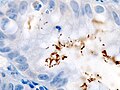
Image:Immunohistochemical_detection_of_Helicobacter_%281%29_histopatholgy.jpg | H. pylori - IHC. (WC) | |||
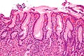
Image:Gastritis_helicobacter_-_high_mag.jpg | Gastritis due to HP. (WC) | |||
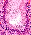
Image:Gastritis_helicobacter_-_very_high_mag_cropped.jpg | HP visible. (WC) | |||
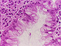
Image:Stomach_helicobacter_he.JPG | HP gastritis. (WC) | |||
Image:Helicobacter_pylori_in_a_case_of_gastritis.jpg | HP gastritis. (WC) | |||
Image:Helicobacter_pylori,_Gastric_Mucosa,_H%26E.jpg | HP gastritis. (WC) | |||
</gallery> | |||
<gallery> | |||
Image: Helicobacter gastritis -- very high mag.jpg | HP - very high mag. (WC) | |||
Image: Helicobacter gastritis -- extremely high mag.jpg | HP - extremely high mag. (WC) | |||
Image: Helicobacter gastritis - crop -- extremely high mag.jpg | HP - extremely high mag. (WC) | |||
Image: Helicobacter gastritis - ext crop -- extremely high mag.jpg | HP - extremely high mag. (WC) | |||
</gallery> | |||
www: | |||
*[http://commons.wikimedia.org/wiki/Category:Helicobacter_gastritis Set of images - HP gastritis (WC)]. | |||
*[http://gut.bmj.com/content/58/12/1669/F2.large.jpg Helicobacter heilmannii (bmj.com)].<ref>URL: [http://gut.bmj.com/content/58/12/1669.extract http://gut.bmj.com/content/58/12/1669.extract]. Accessed on: 2 March 2012.</ref> | |||
====Helicobacter heilmannii==== | |||
<gallery> | |||
File:Helicobacter-heilmannii.JPG | HH. (WC/Patho) | |||
File:Helicobacter-heilmannii_2.JPG | HH. (WC/Patho) | |||
File:Helicobacter-heilmannii_3.JPG | HH. (WC/Patho) | |||
</gallery> | |||
==Stains== | |||
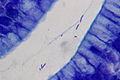
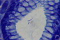
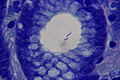
*[[Cresyl violet stain]] - background and organisms blue. | |||
*[[Warthin-Starry stain]] - background yellow, organisms black. | |||
==IHC== | |||
*Helicobacter pylori IHC stain +ve. | |||
Note: | |||
*Reportly also stains ''Helicobacter heilmannii''.<ref name=pmid16224223 >{{Cite journal | last1 = Singhal | first1 = AV. | last2 = Sepulveda | first2 = AR. | title = Helicobacter heilmannii gastritis: a case study with review of literature. | journal = Am J Surg Pathol | volume = 29 | issue = 11 | pages = 1537-9 | month = Nov | year = 2005 | doi = | PMID = 16224223 }}</ref> | |||
==Sign out== | |||
===Antrum=== | |||
<pre> | |||
Stomach, Antrum, Biopsy: | |||
- Antral-type gastric mucosa with abundant HELICOBACTER-LIKE ORGANISMS | |||
and moderate chronic active inflammation. | |||
- NEGATIVE for intestinal metaplasia. | |||
- NEGATIVE for dysplasia and NEGATIVE for malignancy. | |||
</pre> | |||
===Body=== | |||
<pre> | |||
Stomach, Body, Biopsy: | |||
- Body-type gastric mucosa with abundant HELICOBACTER-LIKE ORGANISMS | |||
and moderate chronic active inflammation. | |||
- NEGATIVE for intestinal metaplasia. | |||
- NEGATIVE for dysplasia and NEGATIVE for malignancy. | |||
</pre> | |||
===IELs in duodenum=== | |||
<pre> | |||
A. Duodenum, Biopsy: | |||
- Small bowel mucosa with increased intraepithelial lymphocytes, villous | |||
architecture and crypt architecture within normal limits, see comment. | |||
- NEGATIVE for acute duodenitis. | |||
- NEGATIVE for dysplasia. | |||
B. Stomach, Biopsy: | |||
- Body-type gastric mucosa with moderate chronic active inflammation | |||
and abundant HELICOBACTER-LIKE ORGANISMS. | |||
- NEGATIVE for intestinal metaplasia. | |||
- NEGATIVE for dysplasia and NEGATIVE for malignancy. | |||
COMMENT: | |||
There are approximately 45 lymphocytes/100 enterocytes. Increased intraepithelial lymphocytes (IELs) is a nonspecific finding. IELs are seen in celiac disease and inflammatory bowel disease; however, may be explained by the Helicobacter-like organisms found in the stomach. Clinical correlation is suggested. | |||
</pre> | |||
=====Alternate comment===== | |||
<pre> | |||
Comment: | |||
Focally, there are approximately 50 lymphocytes/100 enterocytes. Increased intraepithelial | |||
lymphocytes is a nonspecific finding that may be seen in a number of conditions, including | |||
infections (e.g. Helicobacter gastritis), and autoimmune disorders (e.g. Crohn's disease, celiac disease). In this case, it may be explained by the Helicobacter-like organisms found in the stomach. Clinical correlation is suggested. | |||
</pre> | |||
====Block letters==== | |||
<pre> | |||
STOMACH, BIOPSY: | |||
- BODY-TYPE GASTRIC MUCOSA WITH MODERATE CHRONIC ACTIVE INFLAMMATION. | |||
- ABUNDANT HELICOBACTER-LIKE ORGANISMS PRESENT. | |||
- NEGATIVE FOR INTESTINAL METAPLASIA. | |||
- NEGATIVE FOR DYSPLASIA AND NEGATIVE FOR MALIGNANCY. | |||
</pre> | |||
===Antrum=== | |||
<pre> | |||
Stomach, Antrum, Biopsy: | |||
- Antral-type gastric mucosa with abundant HELICOBACTER-LIKE ORGANISMS, | |||
and moderate chronic active inflammation. | |||
- NEGATIVE for intestinal metaplasia. | |||
- NEGATIVE for dysplasia and NEGATIVE for malignancy. | |||
</pre> | |||
=====Alternate===== | |||
<pre> | |||
Stomach, Biopsy: | |||
- Antral-type gastric mucosa with moderate chronic active inflammation. | |||
- Abundant Helicobacter-like organisms present. | |||
- NEGATIVE for intestinal metaplasia. | |||
- NEGATIVE for dysplasia and NEGATIVE for malignancy. | |||
</pre> | |||
=====Block letters===== | |||
<pre> | |||
STOMACH, BIOPSY: | |||
- ANTRAL-TYPE GASTRIC MUCOSA WITH MODERATE CHRONIC ACTIVE INFLAMMATION. | |||
- ABUNDANT HELICOBACTER-LIKE ORGANISMS PRESENT. | |||
- NEGATIVE FOR INTESTINAL METAPLASIA. | |||
- NEGATIVE FOR DYSPLASIA AND NEGATIVE FOR MALIGNANCY. | |||
</pre> | |||
====IM present==== | |||
<pre> | |||
Stomach, Biopsy: | |||
- Antral-type gastric mucosa with: | |||
-- Focal intestinal metaplasia. | |||
-- Abundant Helicobacter-like organisms. | |||
-- Moderate chronic active gastritis. | |||
- NEGATIVE for dysplasia and NEGATIVE for malignancy. | |||
</pre> | |||
======Block letters====== | |||
<pre> | |||
STOMACH, BIOPSY: | |||
- ANTRAL-TYPE GASTRIC MUCOSA WITH: | |||
-- FOCAL INTESTINAL METAPLASIA. | |||
-- ABUNDANT HELICOBACTER-LIKE ORGANISMS. | |||
-- MODERATE CHRONIC ACTIVE INFLAMMATION. | |||
- NEGATIVE FOR DYSPLASIA AND NEGATIVE FOR MALIGNANCY. | |||
</pre> | |||
====H. heilmannii==== | |||
<pre> | |||
Stomach, Biopsy: | |||
- Antral-type gastric mucosa with moderate chronic active inflammation. | |||
- Abundant Helicobacter-like organisms present. | |||
- NEGATIVE for intestinal metaplasia. | |||
- NEGATIVE for dysplasia and NEGATIVE for malignancy. | |||
Comment: | |||
The Helicobacter-like organisms have an appearance similar to crinkle cut French fries; this suggests the organisms are H. heilmannii. | |||
</pre> | |||
===Micro=== | |||
The sections show antral-type gastric mucosa with abundant lamina propria plasma cells and | |||
focal intraepithelial neutrophils. Cocci and bacilli are present. Some of the bacilli | |||
are Helicobactor-like. The epithelium matures normally to the surface. No goblet cells | |||
are identified. | |||
==See also== | |||
*[[Stomach]]. | |||
*[[Chronic gastritis]]. | |||
*[[Helicobacter duodenitis]]. | |||
==References== | |||
{{Reflist|2}} | |||
[[Category:Diagnosis]] | [[Category:Diagnosis]] | ||
[[Category:Stomach]] | |||
[[Category:Gastrointestinal pathology]] | |||
Latest revision as of 18:43, 5 February 2024
| Helicobacter gastritis | |
|---|---|
| Diagnosis in short | |
 Helicobacter gastritis. H&E stain. | |
|
| |
| LM | helicobacter organisms, moderate chronic active gastritis (neutrophils esp. at the luminal aspect/intraepithelial, numerous plasma cell clusters) |
| Subtypes | Helicobacter pylori, Helicobacter heilmannii |
| LM DDx | acute gastritis, chronic gastritis |
| Stains | Diff-Quik, Cresyl violet stain, Warthin-Starry stain |
| IHC | Helicobacter IHC |
| Site | stomach |
|
| |
| Associated Dx | MALT lymphoma, gastric carcinoma, intestinal metaplasia of the stomach, peptic ulcer, duodenitis |
| Prevalence | common |
| Endoscopy | erythema |
| Prognosis | benign |
| Clin. DDx | normal stomach |
Helicobacter gastritis, abbreviated HG, is a common form of gastritis caused by Helicobacter species.
The most common Helicobacter implicated is Helicobacter pylori, abbreviated HP.
General
- Several Helicobacter species can cause gastritis:
- Helicobacter pylori - most common.
- Helicobacter heilmannii.
Epidemiologic associations - Helicobacter infections are associated with:[1]
- Gastritis.
- Peptic ulcers.
- Cancer.
- Carcinoma.
- MALT lymphoma.
Note:
- Historically, Helicobacter was grouped with Campylobacter.[2]
- This is why it is the rapid urease test, sometimes done at endoscopy, is also known as the Campylobacter-like organism test, abbreviated CLO test.
Gross
- Thickened gastric folds.
- Erythema.
Microscopic
Features:
- Helicobacter organisms - key feature.
- Inflammation - usually moderate chronic active.
- Clusters of (lamina propria) plasma cells.
- Neutrophils, numerous, classically intraepithelial and superficial.
Tips:
- One needs to look at 400x magnification. Even at 400x they are possible to miss.
- Helicobacter are damn small. They are smaller than the nucleus of the gastric foveollar cell.
- Look for mucus - they preferentially reside there.
- This is usually close to the opening of the gastric pits.
- Helicobacter are found in groups. When you see several that are the same size and shape you can be sure they are real.
Notes:
- Helicobacter can be in antrum and/or body.[5]
- Helicobacter don't like the intestinal mucosa or mucosa that has undergone intestinal metaplasia; you're less likely to find 'em adjacent to it. In general, Helicobacter is uncommon in the context of a case with IM... but common enough that one still ought to look for it.
- May be associated with G-cell hyperplasia.[6]
DDx:
- Dirt - material has a variable size.
- Contamination from oropharynx - bacilli straight, not associated with gastric mucosa.
- Chronic gastritis.
- Acute gastritis.
Images
H. pylori - IHC. (WC)
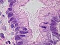
Gastritis due to HP. (WC)
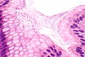
HP visible. (WC)
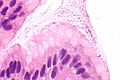
HP gastritis. (WC)
HP gastritis. (WC)
HP gastritis. (WC)


.jpg)

HP - very high mag. (WC)
HP - extremely high mag. (WC)
HP - extremely high mag. (WC)
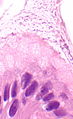
HP - extremely high mag. (WC)


www:
Helicobacter heilmannii
HH. (WC/Patho)
HH. (WC/Patho)
HH. (WC/Patho)


Stains
- Cresyl violet stain - background and organisms blue.
- Warthin-Starry stain - background yellow, organisms black.
IHC
- Helicobacter pylori IHC stain +ve.
Note:
- Reportly also stains Helicobacter heilmannii.[4]
Sign out
Antrum
Stomach, Antrum, Biopsy:
	- Antral-type gastric mucosa with abundant HELICOBACTER-LIKE ORGANISMS
	  and moderate chronic active inflammation.
	- NEGATIVE for intestinal metaplasia.
	- NEGATIVE for dysplasia and NEGATIVE for malignancy.
Body
Stomach, Body, Biopsy:
	- Body-type gastric mucosa with abundant HELICOBACTER-LIKE ORGANISMS
	  and moderate chronic active inflammation.
	- NEGATIVE for intestinal metaplasia.
	- NEGATIVE for dysplasia and NEGATIVE for malignancy.
IELs in duodenum
A. Duodenum, Biopsy:
- Small bowel mucosa with increased intraepithelial lymphocytes, villous
  architecture and crypt architecture within normal limits, see comment.
- NEGATIVE for acute duodenitis.
- NEGATIVE for dysplasia.

B. Stomach, Biopsy:
- Body-type gastric mucosa with moderate chronic active inflammation
  and abundant HELICOBACTER-LIKE ORGANISMS.
- NEGATIVE for intestinal metaplasia.
- NEGATIVE for dysplasia and NEGATIVE for malignancy.

COMMENT:
There are approximately 45 lymphocytes/100 enterocytes. Increased intraepithelial lymphocytes (IELs) is a nonspecific finding. IELs are seen in celiac disease and inflammatory bowel disease; however, may be explained by the Helicobacter-like organisms found in the stomach. Clinical correlation is suggested.
Alternate comment
Comment:
Focally, there are approximately 50 lymphocytes/100 enterocytes. Increased intraepithelial
lymphocytes is a nonspecific finding that may be seen in a number of conditions, including
infections (e.g. Helicobacter gastritis), and autoimmune disorders (e.g. Crohn's disease, celiac disease). In this case, it may be explained by the Helicobacter-like organisms found in the stomach. Clinical correlation is suggested.
Block letters
STOMACH, BIOPSY:
- BODY-TYPE GASTRIC MUCOSA WITH MODERATE CHRONIC ACTIVE INFLAMMATION.
- ABUNDANT HELICOBACTER-LIKE ORGANISMS PRESENT.
- NEGATIVE FOR INTESTINAL METAPLASIA.
- NEGATIVE FOR DYSPLASIA AND NEGATIVE FOR MALIGNANCY.
Antrum
Stomach, Antrum, Biopsy:
     - Antral-type gastric mucosa with abundant HELICOBACTER-LIKE ORGANISMS,
       and moderate chronic active inflammation.
     - NEGATIVE for intestinal metaplasia.
     - NEGATIVE for dysplasia and NEGATIVE for malignancy.
Alternate
Stomach, Biopsy:
- Antral-type gastric mucosa with moderate chronic active inflammation.
- Abundant Helicobacter-like organisms present.
- NEGATIVE for intestinal metaplasia.
- NEGATIVE for dysplasia and NEGATIVE for malignancy.
Block letters
STOMACH, BIOPSY:
- ANTRAL-TYPE GASTRIC MUCOSA WITH MODERATE CHRONIC ACTIVE INFLAMMATION.
- ABUNDANT HELICOBACTER-LIKE ORGANISMS PRESENT.
- NEGATIVE FOR INTESTINAL METAPLASIA.
- NEGATIVE FOR DYSPLASIA AND NEGATIVE FOR MALIGNANCY.
IM present
Stomach, Biopsy:
- Antral-type gastric mucosa with:
-- Focal intestinal metaplasia.
-- Abundant Helicobacter-like organisms.
-- Moderate chronic active gastritis.
- NEGATIVE for dysplasia and NEGATIVE for malignancy.
Block letters
STOMACH, BIOPSY:
- ANTRAL-TYPE GASTRIC MUCOSA WITH:
-- FOCAL INTESTINAL METAPLASIA.
-- ABUNDANT HELICOBACTER-LIKE ORGANISMS.
-- MODERATE CHRONIC ACTIVE INFLAMMATION.
- NEGATIVE FOR DYSPLASIA AND NEGATIVE FOR MALIGNANCY.
H. heilmannii
Stomach, Biopsy:
- Antral-type gastric mucosa with moderate chronic active inflammation.
- Abundant Helicobacter-like organisms present.
- NEGATIVE for intestinal metaplasia.
- NEGATIVE for dysplasia and NEGATIVE for malignancy.

Comment:
The Helicobacter-like organisms have an appearance similar to crinkle cut French fries; this suggests the organisms are H. heilmannii.
Micro
The sections show antral-type gastric mucosa with abundant lamina propria plasma cells and focal intraepithelial neutrophils. Cocci and bacilli are present. Some of the bacilli are Helicobactor-like. The epithelium matures normally to the surface. No goblet cells are identified.
See also
References
- ↑ Cotran, Ramzi S.; Kumar, Vinay; Fausto, Nelson; Nelso Fausto; Robbins, Stanley L.; Abbas, Abul K. (2005). Robbins and Cotran pathologic basis of disease (7th ed.). St. Louis, Mo: Elsevier Saunders. pp. 814. ISBN 0-7216-0187-1.
- ↑ Ciortescu, I.; Stan, M.. "[Helicobacter pylori--friend or foe?].". Rev Med Chir Soc Med Nat Iasi 114 (3): 619-24. PMID 21243784.
- ↑ Mobley, HLT.; Mendz, GL.; Hazell, SL.; Andersen, LP.; Wadström, T.. Basic Bacteriology and Culture. PMID 21290743. http://www.ncbi.nlm.nih.gov/books/NBK2444/.
- ↑ 4.0 4.1 Singhal, AV.; Sepulveda, AR. (Nov 2005). "Helicobacter heilmannii gastritis: a case study with review of literature.". Am J Surg Pathol 29 (11): 1537-9. PMID 16224223.
- ↑ Maaroos HI, Kekki M, Villako K, Sipponen P, Tamm A, Sadeniemi L (October 1990). "The occurrence and extent of Helicobacter pylori colonization and antral and body gastritis profiles in an Estonian population sample". Scand. J. Gastroenterol. 25 (10): 1010-7. PMID 2263873.
- ↑ Kwan, CP.; Tytgat, GN. (Nov 1995). "Antral G-cell hyperplasia: a vanishing disease?". Eur J Gastroenterol Hepatol 7 (11): 1099-1103. PMID 8680911.
- ↑ URL: http://gut.bmj.com/content/58/12/1669.extract. Accessed on: 2 March 2012.