Difference between revisions of "Kidney"
Jump to navigation
Jump to search
(→Developmental: more) |
|||
| (12 intermediate revisions by the same user not shown) | |||
| Line 1: | Line 1: | ||
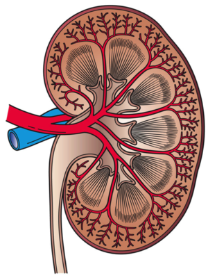
[[Image:Kidney_Cross_Section.png|thumb|right|A drawing of the kidney in cross section. (WC/Holly Fischer)]] | |||
The '''kidney''' is an important organ in the abdomen that does the following: | The '''kidney''' is an important organ in the abdomen that does the following: | ||
*Water balance & blood pressure regulation, | *Water balance & blood pressure regulation, | ||
| Line 4: | Line 5: | ||
*Removes toxins/cleans the blood, and | *Removes toxins/cleans the blood, and | ||
*Produces hormones (e.g. erythropoietin). | *Produces hormones (e.g. erythropoietin). | ||
=Normal= | |||
===Sign out=== | |||
====Missed biopsy==== | |||
<pre> | |||
KIDNEY, RIGHT, BIOPSY: | |||
- SCANT BENIGN ADIPOSE TISSUE AND A FEW SMALL BLOOD VESSELS. | |||
- NO RENAL PARENCHYMA IS IDENTIFIED. | |||
- NO EVIDENCE OF MALIGNANCY. | |||
COMMENT: | |||
The indication (mass lesion) and clinical suspicion is noted. The tissue | |||
sample may not be representative of the lesions seen radiologically. | |||
Clinical correlation is required. | |||
</pre> | |||
=====Micro===== | |||
The sections show benign adipose tissue with a few small blood vessels. The core length (at microscopy) is approximately 9 millimetres. No renal parenchyma is identified. No nuclear atypia is identified. No significant amount of smooth muscle is apparent. | |||
=Tumours= | =Tumours= | ||
| Line 10: | Line 30: | ||
This article cover the common renal tumours: | This article cover the common renal tumours: | ||
*[[Renal cell carcinoma]. | *[[Renal cell carcinoma]]. | ||
*[[Renal oncocytoma|Oncocytoma]]. | *[[Renal oncocytoma|Oncocytoma]]. | ||
*Most other tumours... except ''urothelial tumours'' are dealt with in the ''[[urothelium]]'' article. | *Most other tumours... except ''urothelial tumours'' are dealt with in the ''[[urothelium]]'' article. | ||
Pediatric tumours are covered in ''[[pediatric | Pediatric tumours are covered in ''[[pediatric kidney tumours]]''. | ||
=Medical kidney diseases= | =Medical kidney diseases= | ||
{{main|Medical kidney diseases}} | {{main|Medical kidney diseases}} | ||
This is almost a specialty for itself. Lots of interaction with nephrologists. | This is almost a specialty for itself. Lots of interaction with nephrologists. Cystic renal disease is dealt with in a separate article called ''[[cystic kidney diseases]]''. | ||
=Other= | |||
==Renal segmental hypoplasia== | |||
{{Main|Renal segmental hypoplasia}} | |||
=Developmental= | =Developmental= | ||
==Horseshoe kidney== | |||
===General=== | |||
*Anatomical variant. | |||
*Prevalence ~1 in 500.<ref name=Ref_Klatt233>{{Ref Klatt|233}}</ref> | |||
===Gross=== | |||
*The inferior poles of the kidneys are joined with one another - have the shape of a horseshoe. | |||
Image: | |||
*[http://library.med.utah.edu/WebPath/RENAHTML/RENAL004.html Horseshoe kidney (utah.edu)]. | |||
==Multicystic renal dysplasia== | ==Multicystic renal dysplasia== | ||
*Abbreviated ''MRD''. | |||
===General=== | ===General=== | ||
*Most common cause of abdominal mass in newborns.<ref name=emed_mrd/> | *Most common cause of abdominal mass in newborns.<ref name=emed_mrd/> | ||
*Subtype of renal dysplasia.<ref name=emed_mrd>URL: [http://emedicine.medscape.com/article/982560-overview http://emedicine.medscape.com/article/982560-overview]. Accessed on: 4 January 2012.</ref> | *Subtype of renal dysplasia.<ref name=emed_mrd>URL: [http://emedicine.medscape.com/article/982560-overview http://emedicine.medscape.com/article/982560-overview]. Accessed on: 4 January 2012.</ref> | ||
*May be unilateral or involve only part of a kidney.<ref name=Ref_Klatt237>{{Ref Klatt|237}}</ref> | |||
===Gross=== | |||
*Kidney has multiple large cysts or differing sizes. | |||
DDx: | |||
*[[ARPKD]] - has less variability of cyst size. | |||
Images: | |||
*[http://library.med.utah.edu/WebPath/RENAHTML/RENAL043.html MRD (utah.edu)]. | |||
*[http://radiology.uchc.edu/eAtlas/GU/551.htm MRD (radiology.uchc.edu)]. | |||
===Microscopic=== | ===Microscopic=== | ||
Features: | Features:<ref name=Ref_Klatt237>{{Ref Klatt|237}}</ref> | ||
* | *Cystic spaces. | ||
* | *Fibrous stroma. | ||
*Islands of cartilage. | |||
Image: | |||
*[http://library.med.utah.edu/WebPath/TUTORIAL/RENCYST/RCYST012.html RMD (utah.edu)]. | |||
==Renal medullary dysplasia== | ==Renal medullary dysplasia== | ||
===General=== | ===General=== | ||
*Associated with: | *Associated with: | ||
**[[ | **[[Beckwith-Wiedemann syndrome]].<ref name=pmid17172498>{{Cite journal | last1 = Dotto | first1 = J. | last2 = Reyes-Múgica | first2 = M. | title = Renal medullary dysplasia is diagnostic of Beckwith-Wiedemann syndrome. | journal = Int J Surg Pathol | volume = 15 | issue = 1 | pages = 60-1 | month = Jan | year = 2007 | doi = 10.1177/1066896906295685 | PMID = 17172498 }}</ref> | ||
**[[Placenta|Placental]] insufficiency.<ref name=pmid19297558>{{Cite journal | last1 = Sparrow | first1 = DB. | last2 = Boyle | first2 = SC. | last3 = Sams | first3 = RS. | last4 = Mazuruk | first4 = B. | last5 = Zhang | first5 = L. | last6 = Moeckel | first6 = GW. | last7 = Dunwoodie | first7 = SL. | last8 = de Caestecker | first8 = MP. | title = Placental insufficiency associated with loss of Cited1 causes renal medullary dysplasia. | journal = J Am Soc Nephrol | volume = 20 | issue = 4 | pages = 777-86 | month = Apr | year = 2009 | doi = 10.1681/ASN.2008050547 | PMID = 19297558 }}</ref> | **[[Placenta|Placental]] insufficiency.<ref name=pmid19297558>{{Cite journal | last1 = Sparrow | first1 = DB. | last2 = Boyle | first2 = SC. | last3 = Sams | first3 = RS. | last4 = Mazuruk | first4 = B. | last5 = Zhang | first5 = L. | last6 = Moeckel | first6 = GW. | last7 = Dunwoodie | first7 = SL. | last8 = de Caestecker | first8 = MP. | title = Placental insufficiency associated with loss of Cited1 causes renal medullary dysplasia. | journal = J Am Soc Nephrol | volume = 20 | issue = 4 | pages = 777-86 | month = Apr | year = 2009 | doi = 10.1681/ASN.2008050547 | PMID = 19297558 }}</ref> | ||
===Microscopic=== | |||
Features:<ref name=pmid17172498/> | |||
*Widely spaced tubules in the medulla of the kidney. | |||
=See also= | =See also= | ||
Latest revision as of 18:35, 19 September 2017
The kidney is an important organ in the abdomen that does the following:
- Water balance & blood pressure regulation,
- Acid-base balance,
- Removes toxins/cleans the blood, and
- Produces hormones (e.g. erythropoietin).
Normal
Sign out
Missed biopsy
KIDNEY, RIGHT, BIOPSY:
- SCANT BENIGN ADIPOSE TISSUE AND A FEW SMALL BLOOD VESSELS.
- NO RENAL PARENCHYMA IS IDENTIFIED.
- NO EVIDENCE OF MALIGNANCY.

COMMENT:
The indication (mass lesion) and clinical suspicion is noted.  The tissue
sample may not be representative of the lesions seen radiologically.
Clinical correlation is required.
Micro
The sections show benign adipose tissue with a few small blood vessels. The core length (at microscopy) is approximately 9 millimetres. No renal parenchyma is identified. No nuclear atypia is identified. No significant amount of smooth muscle is apparent.
Tumours
Main article: Kidney tumours
This is mostly the domain of urology.
This article cover the common renal tumours:
- Renal cell carcinoma.
- Oncocytoma.
- Most other tumours... except urothelial tumours are dealt with in the urothelium article.
Pediatric tumours are covered in pediatric kidney tumours.
Medical kidney diseases
Main article: Medical kidney diseases
This is almost a specialty for itself. Lots of interaction with nephrologists. Cystic renal disease is dealt with in a separate article called cystic kidney diseases.
Other
Renal segmental hypoplasia
Main article: Renal segmental hypoplasia
Developmental
Horseshoe kidney
General
- Anatomical variant.
- Prevalence ~1 in 500.[1]
Gross
- The inferior poles of the kidneys are joined with one another - have the shape of a horseshoe.
Image:
Multicystic renal dysplasia
- Abbreviated MRD.
General
- Most common cause of abdominal mass in newborns.[2]
- Subtype of renal dysplasia.[2]
- May be unilateral or involve only part of a kidney.[3]
Gross
- Kidney has multiple large cysts or differing sizes.
DDx:
- ARPKD - has less variability of cyst size.
Images:
Microscopic
Features:[3]
- Cystic spaces.
- Fibrous stroma.
- Islands of cartilage.
Image:
Renal medullary dysplasia
General
- Associated with:
- Beckwith-Wiedemann syndrome.[4]
- Placental insufficiency.[5]
Microscopic
Features:[4]
- Widely spaced tubules in the medulla of the kidney.
See also
References
- ↑ Klatt, Edward C. (2006). Robbins and Cotran Atlas of Pathology (1st ed.). Saunders. pp. 233. ISBN 978-1416002741.
- ↑ 2.0 2.1 URL: http://emedicine.medscape.com/article/982560-overview. Accessed on: 4 January 2012.
- ↑ 3.0 3.1 Klatt, Edward C. (2006). Robbins and Cotran Atlas of Pathology (1st ed.). Saunders. pp. 237. ISBN 978-1416002741.
- ↑ 4.0 4.1 Dotto, J.; Reyes-Múgica, M. (Jan 2007). "Renal medullary dysplasia is diagnostic of Beckwith-Wiedemann syndrome.". Int J Surg Pathol 15 (1): 60-1. doi:10.1177/1066896906295685. PMID 17172498.
- ↑ Sparrow, DB.; Boyle, SC.; Sams, RS.; Mazuruk, B.; Zhang, L.; Moeckel, GW.; Dunwoodie, SL.; de Caestecker, MP. (Apr 2009). "Placental insufficiency associated with loss of Cited1 causes renal medullary dysplasia.". J Am Soc Nephrol 20 (4): 777-86. doi:10.1681/ASN.2008050547. PMID 19297558.
