Difference between revisions of "Peliosis hepatis"
Jump to navigation
Jump to search
| Line 5: | Line 5: | ||
*Rarely biopsied. | *Rarely biopsied. | ||
*May cause hemoperitoneum.<ref name=pmid26675327/><ref name=pmid22104329>{{Cite journal | last1 = Buelow | first1 = B. | last2 = Otjen | first2 = J. | last3 = Sabath | first3 = AP. | last4 = Harruff | first4 = RC. | title = Peliosis hepatis presenting as liver rupture in a vulnerable adult: a case report. | journal = Am J Forensic Med Pathol | volume = 33 | issue = 4 | pages = 307-10 | month = Dec | year = 2012 | doi = 10.1097/PAF.0b013e31823a8b38 | PMID = 22104329 }}</ref> | *May cause hemoperitoneum.<ref name=pmid26675327/><ref name=pmid22104329>{{Cite journal | last1 = Buelow | first1 = B. | last2 = Otjen | first2 = J. | last3 = Sabath | first3 = AP. | last4 = Harruff | first4 = RC. | title = Peliosis hepatis presenting as liver rupture in a vulnerable adult: a case report. | journal = Am J Forensic Med Pathol | volume = 33 | issue = 4 | pages = 307-10 | month = Dec | year = 2012 | doi = 10.1097/PAF.0b013e31823a8b38 | PMID = 22104329 }}</ref> | ||
*Etiology - unknown.<ref name=pmid24137445>{{Cite journal | last1 = Dai | first1 = W. | last2 = Zhong | first2 = D. | title = Peliosis hepatis mimicking cancer: A case report. | journal = Oncol Lett | volume = 6 | issue = 4 | pages = 960-962 | month = Oct | year = 2013 | doi = 10.3892/ol.2013.1479 | PMID = 24137445 }}</ref> | |||
Associated with: | Associated with: | ||
Revision as of 03:37, 19 June 2017
Peliosis hepatis is a type of medical liver disease.
General
Associated with:
- Infections.
- Malignancy.
- Other stuff.
Microscopic
Features:
- Multiple small cysts[1] lined by endothelium.
- Usually incomplete.
- Blood.
Images
A. 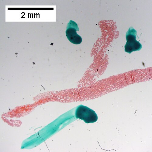
B. 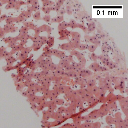
C. 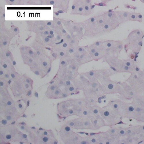
D. 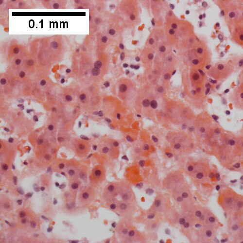
Peliosis hepatis. A. Hemorrhage at left end, dilated sinusoids elsewhere. B. Ramifying dilated sinusoidal spaces. C. PAS with diastase shows flat lining. D. Necrotic hepatocytes in cords, presumably due to pressure.
See also
References
- ↑ 1.0 1.1 1.2 Crocetti, D.; Palmieri, A.; Pedullà, G.; Pasta, V.; D'Orazi, V.; Grazi, GL. (Dec 2015). "Peliosis hepatis: Personal experience and literature review.". World J Gastroenterol 21 (46): 13188-94. doi:10.3748/wjg.v21.i46.13188. PMID 26675327.
- ↑ Buelow, B.; Otjen, J.; Sabath, AP.; Harruff, RC. (Dec 2012). "Peliosis hepatis presenting as liver rupture in a vulnerable adult: a case report.". Am J Forensic Med Pathol 33 (4): 307-10. doi:10.1097/PAF.0b013e31823a8b38. PMID 22104329.
- ↑ Dai, W.; Zhong, D. (Oct 2013). "Peliosis hepatis mimicking cancer: A case report.". Oncol Lett 6 (4): 960-962. doi:10.3892/ol.2013.1479. PMID 24137445.