Difference between revisions of "Hematoxylin and eosin stain"
Jump to navigation
Jump to search
(fix typo found by AC) |
|||
| (One intermediate revision by the same user not shown) | |||
| Line 15: | Line 15: | ||
| Other = | | Other = | ||
}} | }} | ||
'''Hematoxylin and eosin stain''', abbreviated '''H&E''', is the most widely used standard stain in [[pathology]]. | '''Hematoxylin and eosin stain''', abbreviated '''H&E''', is the most widely used standard [[stain]] in [[pathology]]. | ||
==General== | ==General== | ||
Latest revision as of 03:50, 14 July 2016
| Hematoxylin and eosin stain | |
|---|---|
| Stain in short | |
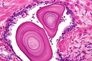
 Hematoxylin and eosin stain of benign prostate. (WC) | |
| Abbreviation | H&E, HE |
| Use | the standard stain in pathology |
| Interpretation | blue (hematoxylin) = nucleus, pink (eosin) = cytoplasm |
Hematoxylin and eosin stain, abbreviated H&E, is the most widely used standard stain in pathology.
General
- Standard bearer in most pathology departments.[1][citation needed]
Intepretation
- Blue (hematoxylin) = nucleus.
- Pink (eosin) = cytoplasm.
Note:
- The above is why it is said (tongue-in-cheek) that blue is bad and pink is dead.
Images
Basal cell carcinoma. H&E stain. (WC)
See also
References
- ↑ Giordano, G. (2009). "Value of immunohistochemistry in uterine pathology: common and rare diagnostic dilemmas.". Pathol Res Pract 205 (10): 663-76. doi:10.1016/j.prp.2009.05.007. PMID 19523774.

