Difference between revisions of "Multifocal micronodular pneumocyte hyperplasia associated with tuberous sclerosis"
Jump to navigation
Jump to search
(tweak) |
|||
| Line 35: | Line 35: | ||
==General== | ==General== | ||
*Rare. | *Rare. | ||
Clinical: | |||
*May present with [[pneumothorax].<ref name=pmid2071093>{{Cite journal | last1 = Popper | first1 = HH. | last2 = Juettner-Smolle | first2 = FM. | last3 = Pongratz | first3 = MG. | title = Micronodular hyperplasia of type II pneumocytes--a new lung lesion associated with tuberous sclerosis. | journal = Histopathology | volume = 18 | issue = 4 | pages = 347-54 | month = Apr | year = 1991 | doi = | PMID = 2071093 }}</ref> | |||
==Gross== | ==Gross== | ||
Revision as of 02:44, 8 March 2016
| Multifocal micronodular pneumocyte hyperplasia associated with tuberous sclerosis | |
|---|---|
| Diagnosis in short | |
 Micronodule of pneumocyte hyperplasia in multifocal micronodular pneumocyte hyperplasia associated with tuberous sclerosis. H&E stain. | |
| LM DDx | atypical adenomatous hyperplasia of the lung |
| Site | lung |
|
| |
| Associated Dx | lymphangioleiomyomatosis - also assoc. with tuberous sclerosis |
| Syndromes | tuberous sclerosis |
|
| |
| Prevalence | rare |
| Prognosis | benign |
Multifocal micronodular pneumocyte hyperplasia associated with tuberous sclerosis, also multifocal micronodular pneumocyte hyperplasia in tuberous sclerosis, is the presence of rare hamartomatous lesions in the lung seen in tuberous sclerosis.[1]
General
- Rare.
Clinical:
- May present with [[pneumothorax].[2]
Gross
Features:[3]
- Multiple small lung nodules - apparently random distribution.
Microscopic
Features:
- Macrophages within the air spaces.
- Enlarged alveolar lining cells with:
- Hobnail morphology - free (luminal) surface area > attached/basal surface area.
- Round or oval nuclei.
DDx:
- Atypical adenomatous hyperplasia of the lung - usu. do not have macrophages within the air spaces.
Images
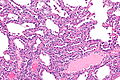
MMPH - intermed. mag.
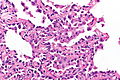
MMPH - high mag.

See also
References
- ↑ Nagar, AM.; Teh, HS.; Khoo, RN.; Morani, AC.; Vrishni, K.; Raghuram, J. (Feb 2008). "Multifocal pneumocyte hyperplasia in tuberous sclerosis.". Thorax 63 (2): 186. doi:10.1136/thx.2006.076604. PMID 18234663.
- ↑ Popper, HH.; Juettner-Smolle, FM.; Pongratz, MG. (Apr 1991). "Micronodular hyperplasia of type II pneumocytes--a new lung lesion associated with tuberous sclerosis.". Histopathology 18 (4): 347-54. PMID 2071093.
- ↑ Kobashi, Y.; Sugiu, T.; Mouri, K.; Irei, T.; Nakata, M.; Oka, M. (Jun 2008). "Multifocal micronodular pneumocyte hyperplasia associated with tuberous sclerosis: differentiation from multiple atypical adenomatous hyperplasia.". Jpn J Clin Oncol 38 (6): 451-4. doi:10.1093/jjco/hyn042. PMID 18535095.