Difference between revisions of "Multifocal micronodular pneumocyte hyperplasia associated with tuberous sclerosis"
Jump to navigation
Jump to search
| Line 1: | Line 1: | ||
'''Multifocal micronodular pneumocyte hyperplasia associated with tuberous sclerosis''', also '''multifocal micronodular pneumocyte hyperplasia in tuberous sclerosis''', is the presence of rare hamartomatous lesions in the [[lung]] seen in [[tuberous sclerosis]].<ref name=pmid18234663 >{{Cite journal | last1 = Nagar | first1 = AM. | last2 = Teh | first2 = HS. | last3 = Khoo | first3 = RN. | last4 = Morani | first4 = AC. | last5 = Vrishni | first5 = K. | last6 = Raghuram | first6 = J. | title = Multifocal pneumocyte hyperplasia in tuberous sclerosis. | journal = Thorax | volume = 63 | issue = 2 | pages = 186 | month = Feb | year = 2008 | doi = 10.1136/thx.2006.076604 | PMID = 18234663 }}</ref> | '''Multifocal micronodular pneumocyte hyperplasia associated with tuberous sclerosis''', also '''multifocal micronodular pneumocyte hyperplasia in tuberous sclerosis''', is the presence of rare hamartomatous lesions in the [[lung]] seen in [[tuberous sclerosis]].<ref name=pmid18234663 >{{Cite journal | last1 = Nagar | first1 = AM. | last2 = Teh | first2 = HS. | last3 = Khoo | first3 = RN. | last4 = Morani | first4 = AC. | last5 = Vrishni | first5 = K. | last6 = Raghuram | first6 = J. | title = Multifocal pneumocyte hyperplasia in tuberous sclerosis. | journal = Thorax | volume = 63 | issue = 2 | pages = 186 | month = Feb | year = 2008 | doi = 10.1136/thx.2006.076604 | PMID = 18234663 }}</ref> | ||
==General== | |||
*Rare. | |||
==Microscopic== | |||
Features: | |||
*Macrophages within the air spaces. | |||
*Enlarged alveolar lining cells with: | |||
**Hobnail morphology - free (luminal) surface area > attached/basal surface area. | |||
**Round or oval nuclei. | |||
DDx: | |||
*[[Atypical adenomatous hyperplasia of the lung]] - usu. do not have macrophages within the air spaces. | |||
===Images=== | |||
<gallery> | |||
Image: Multifocal micronodular pneumocyte hyperplasia - tuberous sclerosis - a3 -- intermed mag.jpg | MMPH - intermed. mag. | |||
Image: Multifocal micronodular pneumocyte hyperplasia - tuberous sclerosis - a3 -- high mag.jpg | MMPH - high mag. | |||
</gallery> | |||
==See also== | ==See also== | ||
Revision as of 01:58, 8 March 2016
Multifocal micronodular pneumocyte hyperplasia associated with tuberous sclerosis, also multifocal micronodular pneumocyte hyperplasia in tuberous sclerosis, is the presence of rare hamartomatous lesions in the lung seen in tuberous sclerosis.[1]
General
- Rare.
Microscopic
Features:
- Macrophages within the air spaces.
- Enlarged alveolar lining cells with:
- Hobnail morphology - free (luminal) surface area > attached/basal surface area.
- Round or oval nuclei.
DDx:
- Atypical adenomatous hyperplasia of the lung - usu. do not have macrophages within the air spaces.
Images
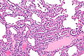
MMPH - intermed. mag.
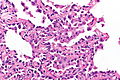
MMPH - high mag.


See also
References
- ↑ Nagar, AM.; Teh, HS.; Khoo, RN.; Morani, AC.; Vrishni, K.; Raghuram, J. (Feb 2008). "Multifocal pneumocyte hyperplasia in tuberous sclerosis.". Thorax 63 (2): 186. doi:10.1136/thx.2006.076604. PMID 18234663.