Difference between revisions of "Focal nodular hyperplasia"
Jump to navigation
Jump to search
(split out) |
(+infobox) |
||
| Line 1: | Line 1: | ||
{{ Infobox diagnosis | |||
| Name = {{PAGENAME}} | |||
| Image = Focal_nodular_hyperplasia_-_intermed_mag.jpg | |||
| Width = | |||
| Caption = Focal nodular hyperplasia. [[H&E stain]]. | |||
| Synonyms = | |||
| Micro = thick walled blood vessels without bile ducts of same size, bile ductular proliferation at the edge of the fibrosis tissue | |||
| Subtypes = | |||
| LMDDx = [[hepatic adenoma]], [[cirrhosis]] | |||
| Stains = | |||
| IHC = | |||
| EM = | |||
| Molecular = | |||
| IF = | |||
| Gross = well circumscribed with capsule, lighter than surrounding parenchyma - may be yellow, +/-stellate central scar with thick vessels | |||
| Grossing = | |||
| Site = [[liver]] - see ''[[medical liver disease]]'' | |||
| Assdx = | |||
| Syndromes = [[hereditary hemorrhagic telangiectasia]] | |||
| Clinicalhx = | |||
| Signs = | |||
| Symptoms = | |||
| Prevalence = | |||
| Bloodwork = | |||
| Rads = usu. solitary lesion, arterial phase enhancement in triphasic imaging | |||
| Endoscopy = | |||
| Prognosis = benign | |||
| Other = | |||
| ClinDDx = | |||
| Tx = | |||
}} | |||
'''Focal nodular hyperplasia''', abbreviated '''FNH''', is a benign [[liver]] lesion, uncommonly seen by pathologists. | '''Focal nodular hyperplasia''', abbreviated '''FNH''', is a benign [[liver]] lesion, uncommonly seen by pathologists. | ||
| Line 17: | Line 48: | ||
*Lighter than surrounding parenchyma, may be yellow. | *Lighter than surrounding parenchyma, may be yellow. | ||
*+/-Stellate central scar with thick vessels. | *+/-Stellate central scar with thick vessels. | ||
**Can be identified on | **Can be identified on medical imaging. | ||
Note: Usually a solitary lesion.<ref name=emedicine_fnh/> | Note: Usually a solitary lesion.<ref name=emedicine_fnh/> | ||
Revision as of 04:45, 17 September 2014
| Focal nodular hyperplasia | |
|---|---|
| Diagnosis in short | |
 Focal nodular hyperplasia. H&E stain. | |
|
| |
| LM | thick walled blood vessels without bile ducts of same size, bile ductular proliferation at the edge of the fibrosis tissue |
| LM DDx | hepatic adenoma, cirrhosis |
| Gross | well circumscribed with capsule, lighter than surrounding parenchyma - may be yellow, +/-stellate central scar with thick vessels |
| Site | liver - see medical liver disease |
|
| |
| Syndromes | hereditary hemorrhagic telangiectasia |
|
| |
| Radiology | usu. solitary lesion, arterial phase enhancement in triphasic imaging |
| Prognosis | benign |
Focal nodular hyperplasia, abbreviated FNH, is a benign liver lesion, uncommonly seen by pathologists.
General
- Not commonly seen by pathologists, as these are usually distinctive on medical imaging.[1]
- Benign lesions.
- May be seen in the context of hereditary hemorrhagic telangiectasia.[2]
Note:
- Oral contraceptive pill (OCP) use does not appear to be a factor in the growth of these lesions;[3] however, the study claims there is nothing on hepatocellular adenomas -- yet I found a JAMA paper by Rooks et al.[4] on this topic.
Imaging
Gross
Features:[6]
- Well circumscribed, but no capsule.
- Lighter than surrounding parenchyma, may be yellow.
- +/-Stellate central scar with thick vessels.
- Can be identified on medical imaging.
Note: Usually a solitary lesion.[5]
Microscopic
Features:[6]
- Classically a stellate scar that has large arteries with fibromuscular hyperplasia.
- Thin fibrous septa radiate from the central scar - surrounded by lymphocytes & bile ductules.
- Normal hepatocytes between fibrous septae.
- Thin fibrous septa radiate from the central scar - surrounded by lymphocytes & bile ductules.
Practical features:
- Thick walled blood vessels.
- Bile duct of same size not seen.
- Bile ductular proliferation at the edge of the fibrosis tissue.
- Clinical history: it is a focal lesion.
DDx:
- Hepatic adenoma - may be difficult to distinguish, if no scar and no ductal proliferation.[7]
- Cirrhosis - complete nodules
- FNH has incomplete nodules.
Memory device FNH = focal lesion, numerous bile ductules, hyperplasia of arteries.
Images
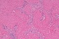
FNH - looks a bit like cirrhosis - low mag. (WC)
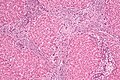
FNH - intermed. mag. (WC)
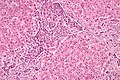
FNH - high mag. (WC)


www:
See also
References
- ↑ 1.0 1.1 Brancatelli, G.; Federle, MP.; Grazioli, L.; Blachar, A.; Peterson, MS.; Thaete, L. (Apr 2001). "Focal nodular hyperplasia: CT findings with emphasis on multiphasic helical CT in 78 patients.". Radiology 219 (1): 61-8. PMID 11274535.
- ↑ Khalid SK, Garcia-Tsao G (August 2008). "Hepatic vascular malformations in hereditary hemorrhagic telangiectasia". Semin. Liver Dis. 28 (3): 247–58. doi:10.1055/s-0028-1085093. PMID 18814078.
- ↑ Kapp, N.; Curtis, KM. (Oct 2009). "Hormonal contraceptive use among women with liver tumors: a systematic review.". Contraception 80 (4): 387-90. doi:10.1016/j.contraception.2009.01.021. PMID 19751862.
- ↑ Rooks, JB.; Ory, HW.; Ishak, KG.; Strauss, LT.; Greenspan, JR.; Hill, AP.; Tyler, CW. (Aug 1979). "Epidemiology of hepatocellular adenoma. The role of oral contraceptive use.". JAMA 242 (7): 644-8. PMID 221698.
- ↑ 5.0 5.1 http://emedicine.medscape.com/article/368377-overview
- ↑ 6.0 6.1 Cotran, Ramzi S.; Kumar, Vinay; Fausto, Nelson; Nelso Fausto; Robbins, Stanley L.; Abbas, Abul K. (2005). Robbins and Cotran pathologic basis of disease (7th ed.). St. Louis, Mo: Elsevier Saunders. pp. 922. ISBN 0-7216-0187-1.
- ↑ STC. 19 Jan 2009.