Difference between revisions of "Angiomatoid fibrous histiocytoma"
Jump to navigation
Jump to search
(+cat.) |
|||
| (5 intermediate revisions by the same user not shown) | |||
| Line 1: | Line 1: | ||
{{ Infobox diagnosis | |||
| Name = {{PAGENAME}} | |||
| Image = Angiomatoid_fibrous_histiocytoma_-_intermed_mag.jpg | |||
| Width = | |||
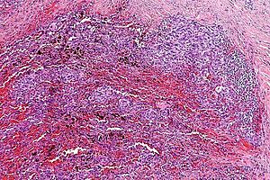
| Caption = Angiomatoid fibrous histiocytoma. [[H&E stain]]. | |||
| Synonyms = | |||
| Micro = cystic spaces with blood +/- histiocytic appearance; inflammation (lymphocytes around periphery of lesion); hemorrhage | |||
| Subtypes = | |||
| LMDDx = | |||
| Stains = | |||
| IHC = CD68 +ve, CD57 +ve, desmin +ve (focal), vimentin +ve | |||
| EM = | |||
| Molecular = t(12;16) FUS/ATF1, t(12;22) EWS/ATF1, others | |||
| IF = | |||
| Gross = | |||
| Grossing = | |||
| Site = [[soft tissue lesions|soft tissue]] - usu. extremities | |||
| Assdx = | |||
| Syndromes = | |||
| Clinicalhx = children, young adults | |||
| Signs = | |||
| Symptoms = | |||
| Prevalence = uncommon | |||
| Bloodwork = | |||
| Rads = | |||
| Endoscopy = | |||
| Prognosis = usually good | |||
| Other = | |||
| ClinDDx = | |||
| Tx = complete excision | |||
}} | |||
'''Angiomatoid fibrous histiocytoma''', abbreviated '''AFH''', is a rare [[soft tissue lesion]] that is typically seen in children and young adults. | |||
==General== | |||
*Rarely metastasizes. | |||
*Children & young adults. | |||
*Should be completely excised. | |||
==Gross== | |||
*Usu. soft tissue of the extremities.{{fact}} | |||
==Microscopic== | |||
Features:<ref name=Ref_WMSP624-5>{{Ref WMSP|624-5}}</ref> | |||
*Cystic spaces with blood - simulates a vascular neoplasm.<ref name=pmid228836>{{Cite journal | last1 = Enzinger | first1 = FM. | title = Angiomatoid malignant fibrous histiocytoma: a distinct fibrohistiocytic tumor of children and young adults simulating a vascular neoplasm. | journal = Cancer | volume = 44 | issue = 6 | pages = 2147-57 | month = Dec | year = 1979 | doi = | PMID = 228836 }}</ref> | |||
*Epithelioid to spindle cells. | |||
**May have a histiocytic appearance.<ref>URL: [http://dermatology.cdlib.org/1605/1_case_reports/4_09-00041/patrizi.html http://dermatology.cdlib.org/1605/1_case_reports/4_09-00041/patrizi.html]. Accessed on: 15 November 2011.</ref> | |||
*Inflammation. | |||
**Lymphoid cuff<ref name=pmid20154033/> - lymphocytes around periphery of lesion. | |||
*Hemorrhage. | |||
Note: | |||
*The first impression may be that it is [[granuloma|granulomatous inflammation]]; however, the cytoplasm doesn't fit (it isn't bubbly and it isn't sheet-like), and the nuclei aren't quite right (few footprint shaped nuclei). | |||
===Images=== | |||
<gallery> | |||
Image: Angiomatoid fibrous histiocytoma - low mag.jpg | AFH - low mag. | |||
Image: Angiomatoid fibrous histiocytoma - intermed mag.jpg | AFH - intermed. mag. | |||
Image: Angiomatoid fibrous histiocytoma - high mag.jpg | AFH - high mag. | |||
Image: Angiomatoid fibrous histiocytoma - very high mag.jpg | AFH - very high mag. | |||
Image: Angiomatoid fibrous histiocytoma - intermed mag - 2.jpg | AFH - intermed. mag. | |||
Image: Angiomatoid fibrous histiocytoma - high mag - 2.jpg | AFH - high mag. | |||
Image: Angiomatoid fibrous histiocytoma - very high mag - 2.jpg | AFH - very high mag. | |||
</gallery> | |||
www: | |||
*[http://www.sarcomaimages.com/index.php?v=Angiomatoid-Fibrous-Histiocytoma AFH (sarcomaimages.com)]. | |||
*[http://jcp.bmj.com/content/63/2/124/F1.large.jpg AFH (bmj.com)].<ref name=pmid20154033>{{Cite journal | last1 = Matsumura | first1 = T. | last2 = Yamaguchi | first2 = T. | last3 = Tochigi | first3 = N. | last4 = Wada | first4 = T. | last5 = Yamashita | first5 = T. | last6 = Hasegawa | first6 = T. | title = Angiomatoid fibrous histiocytoma including cases with pleomorphic features analysed by fluorescence in situ hybridisation. | journal = J Clin Pathol | volume = 63 | issue = 2 | pages = 124-8 | month = Feb | year = 2010 | doi = 10.1136/jcp.2009.072256 | PMID = 20154033 | url = http://jcp.bmj.com/content/63/2/124.full }} | |||
</ref> | |||
*[http://path.upmc.edu/cases/case512.html AFH - several images (upmc.edu)]. | |||
==IHC== | |||
Features:<ref name=Ref_WMSP624-5>{{Ref WMSP|624-5}}</ref> | |||
*CD68 +ve. | |||
*CD57 +ve. | |||
*Desmin +ve (focal). | |||
*Vimentin +ve. | |||
==Molecular== | |||
AFH has recurrent [[translocations]]: | |||
*t(12;16) FUS/ATF1. | |||
*t(12;22) EWS/ATF1. | |||
==See also== | |||
*[[Soft tissue lesions]]. | |||
==References== | |||
{{Reflist|2}} | |||
[[Category:Diagnosis]] | [[Category:Diagnosis]] | ||
[[Category:Soft tissue lesions]] | |||
[[Category:Pediatric pathology]] | |||
Latest revision as of 02:53, 22 June 2014
| Angiomatoid fibrous histiocytoma | |
|---|---|
| Diagnosis in short | |
 Angiomatoid fibrous histiocytoma. H&E stain. | |
|
| |
| LM | cystic spaces with blood +/- histiocytic appearance; inflammation (lymphocytes around periphery of lesion); hemorrhage |
| IHC | CD68 +ve, CD57 +ve, desmin +ve (focal), vimentin +ve |
| Molecular | t(12;16) FUS/ATF1, t(12;22) EWS/ATF1, others |
| Site | soft tissue - usu. extremities |
|
| |
| Clinical history | children, young adults |
| Prevalence | uncommon |
| Prognosis | usually good |
| Treatment | complete excision |
Angiomatoid fibrous histiocytoma, abbreviated AFH, is a rare soft tissue lesion that is typically seen in children and young adults.
General
- Rarely metastasizes.
- Children & young adults.
- Should be completely excised.
Gross
- Usu. soft tissue of the extremities.[citation needed]
Microscopic
Features:[1]
- Cystic spaces with blood - simulates a vascular neoplasm.[2]
- Epithelioid to spindle cells.
- May have a histiocytic appearance.[3]
- Inflammation.
- Lymphoid cuff[4] - lymphocytes around periphery of lesion.
- Hemorrhage.
Note:
- The first impression may be that it is granulomatous inflammation; however, the cytoplasm doesn't fit (it isn't bubbly and it isn't sheet-like), and the nuclei aren't quite right (few footprint shaped nuclei).
Images
www:
IHC
Features:[1]
- CD68 +ve.
- CD57 +ve.
- Desmin +ve (focal).
- Vimentin +ve.
Molecular
AFH has recurrent translocations:
- t(12;16) FUS/ATF1.
- t(12;22) EWS/ATF1.
See also
References
- ↑ 1.0 1.1 Humphrey, Peter A; Dehner, Louis P; Pfeifer, John D (2008). The Washington Manual of Surgical Pathology (1st ed.). Lippincott Williams & Wilkins. pp. 624-5. ISBN 978-0781765275.
- ↑ Enzinger, FM. (Dec 1979). "Angiomatoid malignant fibrous histiocytoma: a distinct fibrohistiocytic tumor of children and young adults simulating a vascular neoplasm.". Cancer 44 (6): 2147-57. PMID 228836.
- ↑ URL: http://dermatology.cdlib.org/1605/1_case_reports/4_09-00041/patrizi.html. Accessed on: 15 November 2011.
- ↑ 4.0 4.1 Matsumura, T.; Yamaguchi, T.; Tochigi, N.; Wada, T.; Yamashita, T.; Hasegawa, T. (Feb 2010). "Angiomatoid fibrous histiocytoma including cases with pleomorphic features analysed by fluorescence in situ hybridisation.". J Clin Pathol 63 (2): 124-8. doi:10.1136/jcp.2009.072256. PMID 20154033. http://jcp.bmj.com/content/63/2/124.full.


