Difference between revisions of "Rheumatoid arthritis"
Jump to navigation
Jump to search
(+joint picture) |
m (rm spc) |
||
| Line 8: | Line 8: | ||
Images: | Images: | ||
*[http://library.med.utah.edu/WebPath/jpeg3/BONE044.jpg RA (med.utah.edu)].<ref>URL: [http://library.med.utah.edu/WebPath/EXAM/IMGQUIZ/msfrm.html http://library.med.utah.edu/WebPath/EXAM/IMGQUIZ/msfrm.html]. Accessed on: 5 December 2010.</ref> | *[http://library.med.utah.edu/WebPath/jpeg3/BONE044.jpg RA (med.utah.edu)].<ref>URL: [http://library.med.utah.edu/WebPath/EXAM/IMGQUIZ/msfrm.html http://library.med.utah.edu/WebPath/EXAM/IMGQUIZ/msfrm.html]. Accessed on: 5 December 2010.</ref> | ||
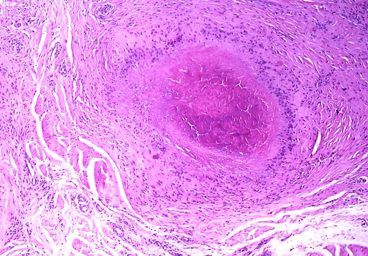
==Rheumatoid nodule== | ==Rheumatoid nodule== | ||
Revision as of 04:09, 6 December 2010
Rheumatoid arthritis, commonly abbreviated RA, is an autoimmune disorder.
Joints
Microscopic
Features:
- Chronic inflammation (e.g. lymphocytes).
Images:
Rheumatoid nodule
General
- Usually only in seropositive cases.[2]
Microscopic
Features:[2]
- Necrobiotic collagen surrounded by:
- Plasma cells.
- Palisading macrophages.
Notes:
- Histomorphologically identical to Granuloma annulare.
Images:
- Rheumatoid nodule (granuloma.homestead.com).[3]
- Rheumatoid nodule (granuloma.homestead.com).[3]
- Rheumatoid nodule (utah.edu).[4]
Lung disease
- See Medical lung disease.
RA may involve the lung.
Amyloidosis
- See Amyloidosis.
Amyloidosis may be due to RA.
See also
References
- ↑ URL: http://library.med.utah.edu/WebPath/EXAM/IMGQUIZ/msfrm.html. Accessed on: 5 December 2010.
- ↑ 2.0 2.1 Tadrous, Paul.J. Diagnostic Criteria Handbook in Histopathology: A Surgical Pathology Vade Mecum (1st ed.). Wiley. pp. 299. ISBN 978-0470519035.
- ↑ 3.0 3.1 URL: http://granuloma.homestead.com/palisading.html. Accessed on: 1 November 2010.
- ↑ URL: http://www.pathguy.com/lectures/joints.htm. Accessed on: 1 November 2010.