Difference between revisions of "Desmoid-type fibromatosis"
Jump to navigation
Jump to search
(+cat.) |
(split-out) |
||
| Line 1: | Line 1: | ||
'''Desmoid-type fibromatosis''' is a benign [[soft tissue lesion]] in the [[fibroblastic/myofibroblastic tumours|fibroblastic/myofibroblastic group of tumours]]. | |||
It is also known as '''desmoid tumour''' and '''desmoid fibromatosis'''. | |||
==General== | |||
*Benign. | |||
*One of many ''[[fibromatoses]]''. | |||
*Locally aggressive.<ref>URL: [http://www.dtrf.org/dtrf_aboutdesmoids.htm http://www.dtrf.org/dtrf_aboutdesmoids.htm]. Accessed on: 15 April 2011.</ref> | |||
*May be seen in the context of [[familial adenomatous polyposis]]. | |||
==Gross== | |||
Features:<ref name=Ref_WMSP609>{{Ref WMSP|609}}</ref> | |||
*Location: | |||
**Abdominal wall, proximal extremities - classic for adolescents and women. | |||
**Head and neck - classic for children. | |||
*Circumscribed mass. | |||
*May be quite large (>10 cm). | |||
==Microscopic== | |||
Features:<ref name=Ref_WMSP609>{{Ref WMSP|609}}</ref><ref>URL: [http://www.surgicalpathologyatlas.com/glfusion/mediagallery/media.php?f=0&sort=0&s=20090717111548196 http://www.surgicalpathologyatlas.com/glfusion/mediagallery/media.php?f=0&sort=0&s=20090717111548196]. Accessed on: 4 October 2011.</ref> | |||
*"Sweeping fascicles"/bundles. | |||
*Spindle cells with: | |||
**Small slender nuclei. | |||
**Solid dark eosinophilic cytoplasm. | |||
*+/-Mitoses - may be abundant. | |||
*Long thin-walled vessels - parallel to one another - '''important feature'''. | |||
DDx: | |||
*[[Hypertrophic scar]]-like lesion, see ''[[dermal scar]]''. | |||
*[[Gastrointestinal stromal tumour]]<ref name=pmid23020601>{{Cite journal | last1 = Huss | first1 = S. | last2 = Nehles | first2 = J. | last3 = Binot | first3 = E. | last4 = Wardelmann | first4 = E. | last5 = Mittler | first5 = J. | last6 = Kleine | first6 = MA. | last7 = Künstlinger | first7 = H. | last8 = Hartmann | first8 = W. | last9 = Hohenberger | first9 = P. | title = β-catenin (CTNNB1) mutations and clinicopathological features of mesenteric desmoid-type fibromatosis. | journal = Histopathology | volume = 62 | issue = 2 | pages = 294-304 | month = Jan | year = 2013 | doi = 10.1111/j.1365-2559.2012.04355.x | PMID = 23020601 }}</ref> | |||
- reported in abdominal wall.<ref name=pmid15257404>{{Cite journal | last1 = Thalheimer | first1 = A. | last2 = Meyer | first2 = D. | last3 = Gattenlöhner | first3 = S. | last4 = Timmermann | first4 = W. | last5 = Thiede | first5 = A. | title = [Gastrointestinal stromal tumor of the abdominal wall. An unusual localization of a rare tumor]. | journal = Chirurg | volume = 75 | issue = 7 | pages = 708-12 | month = Jul | year = 2004 | doi = 10.1007/s00104-003-0696-5 | PMID = 15257404 }}</ref> | |||
*[[Retroperitoneal fibrosis]] - no beta-catenin staining.<ref name=pmid23020601/> | |||
*Other [[fibromatoses]]. | |||
*[[Nodular fasciitis]] - esp. with [[RBC extravasation]]. | |||
===Images=== | |||
<gallery> | |||
Image:DesmoidFibromatosis.JPG | Desmoid tumour. (WC) | |||
</gallery> | |||
www: | |||
*[http://www.surgicalpathologyatlas.com/glfusion/mediagallery/media.php?f=0&sort=0&s=20090717111548196 Desmoid tumour (surgicalpathologyatlas.com)]. | |||
*[http://www.cheapmedicinechest.com/wp-content/uploads/2010/10/Figure-4.-Desmoid-tumor-with-fibroblastic-proliferation.png Desmoid tumour (cheapmedicinechest.com)].<ref>URL: [http://www.cheapmedicinechest.com/abdominal-pain-and-colonic-obstruction-from-an-intra-abdominal-desmoid-tumor.html http://www.cheapmedicinechest.com/abdominal-pain-and-colonic-obstruction-from-an-intra-abdominal-desmoid-tumor.html]. Accessed on: 4 October 2011.</ref> | |||
*[http://radiographics.rsna.org/content/29/7/2143/F28.expansion.html Desmoid tumour (radiographics.rsna.org)]. | |||
*[http://www.ncbi.nlm.nih.gov.qe2a-proxy.mun.ca/pmc/articles/PMC3700980/figure/f3-ol-05-06-1976/ Desmoid-type fibromatosis (nih.gov)].<ref name=pmid23833679>{{Cite journal | last1 = Ma | first1 = JH. | last2 = Ma | first2 = ZH. | last3 = Dong | first3 = XF. | last4 = Yin | first4 = H. | last5 = Zhao | first5 = YF. | title = Abdominal wall desmoid tumors: A case report. | journal = Oncol Lett | volume = 5 | issue = 6 | pages = 1976-1978 | month = Jun | year = 2013 | doi = 10.3892/ol.2013.1297 | PMID = 23833679 }}</ref> | |||
==IHC== | |||
Features:<ref name=Ref_WMSP609>{{Ref WMSP|609}}</ref> | |||
*Beta-catenin +ve (nuclear<ref name=pmid23020601/>) - '''important'''. | |||
*SMA +ve ~50% of lesions. | |||
Others: | |||
*CD117 -ve. | |||
==Sign out== | |||
<pre> | |||
LESION, ABDOMINAL WALL, BIOPSY: | |||
- DESMOID-TYPE FIBROMATOSIS. | |||
COMMENT: | |||
The tumour stains strongly with beta-catenin and weakly with SMA. It is negative for CD117. | |||
</pre> | |||
==See also== | |||
*[[Fibroblastic/myofibroblastic tumours]]. | |||
==References== | |||
{{Reflist|2}} | |||
[[Category:Fibroblastic/myofibroblastic tumours]] | |||
[[Category:Diagnosis]] | [[Category:Diagnosis]] | ||
Revision as of 00:07, 9 November 2013
Desmoid-type fibromatosis is a benign soft tissue lesion in the fibroblastic/myofibroblastic group of tumours.
It is also known as desmoid tumour and desmoid fibromatosis.
General
- Benign.
- One of many fibromatoses.
- Locally aggressive.[1]
- May be seen in the context of familial adenomatous polyposis.
Gross
Features:[2]
- Location:
- Abdominal wall, proximal extremities - classic for adolescents and women.
- Head and neck - classic for children.
- Circumscribed mass.
- May be quite large (>10 cm).
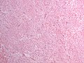
Microscopic
- "Sweeping fascicles"/bundles.
- Spindle cells with:
- Small slender nuclei.
- Solid dark eosinophilic cytoplasm.
- +/-Mitoses - may be abundant.
- Long thin-walled vessels - parallel to one another - important feature.
DDx:
- Hypertrophic scar-like lesion, see dermal scar.
- Gastrointestinal stromal tumour[4]
- reported in abdominal wall.[5]
- Retroperitoneal fibrosis - no beta-catenin staining.[4]
- Other fibromatoses.
- Nodular fasciitis - esp. with RBC extravasation.
Images
Desmoid tumour. (WC)

www:
- Desmoid tumour (surgicalpathologyatlas.com).
- Desmoid tumour (cheapmedicinechest.com).[6]
- Desmoid tumour (radiographics.rsna.org).
- Desmoid-type fibromatosis (nih.gov).[7]
IHC
Features:[2]
- Beta-catenin +ve (nuclear[4]) - important.
- SMA +ve ~50% of lesions.
Others:
- CD117 -ve.
Sign out
LESION, ABDOMINAL WALL, BIOPSY:
- DESMOID-TYPE FIBROMATOSIS.

COMMENT:
The tumour stains strongly with beta-catenin and weakly with SMA. It is negative for CD117.
See also
References
- ↑ URL: http://www.dtrf.org/dtrf_aboutdesmoids.htm. Accessed on: 15 April 2011.
- ↑ 2.0 2.1 2.2 Humphrey, Peter A; Dehner, Louis P; Pfeifer, John D (2008). The Washington Manual of Surgical Pathology (1st ed.). Lippincott Williams & Wilkins. pp. 609. ISBN 978-0781765275.
- ↑ URL: http://www.surgicalpathologyatlas.com/glfusion/mediagallery/media.php?f=0&sort=0&s=20090717111548196. Accessed on: 4 October 2011.
- ↑ 4.0 4.1 4.2 Huss, S.; Nehles, J.; Binot, E.; Wardelmann, E.; Mittler, J.; Kleine, MA.; Künstlinger, H.; Hartmann, W. et al. (Jan 2013). "β-catenin (CTNNB1) mutations and clinicopathological features of mesenteric desmoid-type fibromatosis.". Histopathology 62 (2): 294-304. doi:10.1111/j.1365-2559.2012.04355.x. PMID 23020601.
- ↑ Thalheimer, A.; Meyer, D.; Gattenlöhner, S.; Timmermann, W.; Thiede, A. (Jul 2004). "[Gastrointestinal stromal tumor of the abdominal wall. An unusual localization of a rare tumor].". Chirurg 75 (7): 708-12. doi:10.1007/s00104-003-0696-5. PMID 15257404.
- ↑ URL: http://www.cheapmedicinechest.com/abdominal-pain-and-colonic-obstruction-from-an-intra-abdominal-desmoid-tumor.html. Accessed on: 4 October 2011.
- ↑ Ma, JH.; Ma, ZH.; Dong, XF.; Yin, H.; Zhao, YF. (Jun 2013). "Abdominal wall desmoid tumors: A case report.". Oncol Lett 5 (6): 1976-1978. doi:10.3892/ol.2013.1297. PMID 23833679.