Difference between revisions of "Oral pathology"
Jump to navigation
Jump to search
(+oral fibroma) |
(→Oral fibroma: tweak) |
||
| Line 8: | Line 8: | ||
===General=== | ===General=== | ||
*Most common oral cavity tumour.<ref name=Ref_HaNP240>{{Ref HaNP|240}}</ref> | *Most common oral cavity tumour.<ref name=Ref_HaNP240>{{Ref HaNP|240}}</ref> | ||
*Female | *Female predominance (female:male = 2:1), usually 30-50 years old.<ref name=Ref_HaNP240>{{Ref HaNP|240}}</ref> | ||
*Multiple oral fibromas may be seen in [[Cowden disease]].<ref name=pmid16878060>{{Cite journal | last1 = Segura Saint-Gerons | first1 = R. | last2 = Ceballos Salobreña | first2 = A. | last3 = Toro Rojas | first3 = M. | last4 = Gándara Rey | first4 = JM. | title = Oral manifestations of Cowden's disease. Presentation of a clinical case. | journal = Med Oral Patol Oral Cir Bucal | volume = 11 | issue = 5 | pages = E421-4 | month = Aug | year = 2006 | doi = | PMID = 16878060 }}</ref> | *Multiple oral fibromas may be seen in [[Cowden disease]].<ref name=pmid16878060>{{Cite journal | last1 = Segura Saint-Gerons | first1 = R. | last2 = Ceballos Salobreña | first2 = A. | last3 = Toro Rojas | first3 = M. | last4 = Gándara Rey | first4 = JM. | title = Oral manifestations of Cowden's disease. Presentation of a clinical case. | journal = Med Oral Patol Oral Cir Bucal | volume = 11 | issue = 5 | pages = E421-4 | month = Aug | year = 2006 | doi = | PMID = 16878060 }}</ref> | ||
Revision as of 15:25, 20 August 2012
Oral pathology is a domain of dentistry. In the context of anatomical pathology, it can be lumped with head and neck pathology.
Odontogenic tumours and cysts
Main article: Odontogenic tumours and cysts
Oral neoplasms
Oral fibroma
General
- Most common oral cavity tumour.[1]
- Female predominance (female:male = 2:1), usually 30-50 years old.[1]
- Multiple oral fibromas may be seen in Cowden disease.[2]
- Histologically similar to fibrous papule.[3]
Microscopic
Features:[3]
- Fibrous stroma.
- +/-Collagen bundles.
- Prominent (dilated) vessels.
- Overlying (squamous) mucosa +/-hyperkeratosis +/-focal ulceration.[1]
Pigmented lesions of the oral cavity
A brief DDx of pigmented lesions:[4]
- Diffuse & bilateral:
- Peutz-Jeghers syndrome.
- Addison's disease.
- Drug-induced.
- Smoker's melanosis.
- Focal:
- Vascular lesions.
- Amalgam tattoo.
- Melanocytic lesions.
- Melanotic macule.
- Blue nevus.
- Malignant melanoma.
Smoker's melanosis
General
- Benign.
- Seen in ~20% of smokers.[4]
- Presence of find (smoking) dose-dependent, i.e. longer heavier smokers are more likely to have it.
Gross
- Typically labial gingvia or buccal mucosa.[4]
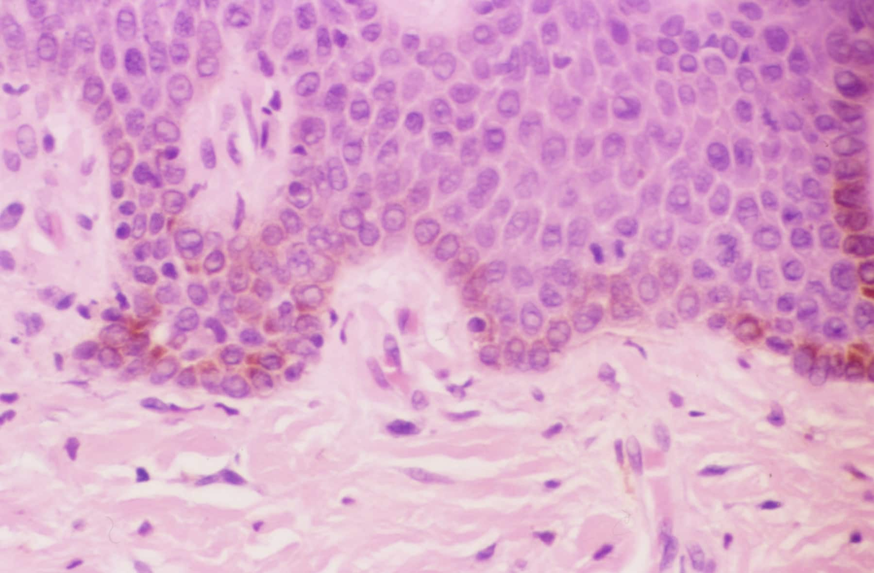
Microscopic
Features:
- Basal melanosis.
- +/-Melanin incontinence.
Image:
See also
References
- ↑ 1.0 1.1 1.2 Thompson, Lester D. R. (2006). Head and Neck Pathology: A Volume in Foundations in Diagnostic Pathology Series (1st ed.). Churchill Livingstone. pp. 240. ISBN 978-0443069604.
- ↑ Segura Saint-Gerons, R.; Ceballos Salobreña, A.; Toro Rojas, M.; Gándara Rey, JM. (Aug 2006). "Oral manifestations of Cowden's disease. Presentation of a clinical case.". Med Oral Patol Oral Cir Bucal 11 (5): E421-4. PMID 16878060.
- ↑ 3.0 3.1 Fernandez-Flores, A. (Jul 2010). "Solitary oral fibromas of the tongue show similar morphologic features to fibrous papule of the face: a study of 31 cases.". Am J Dermatopathol 32 (5): 442-7. doi:10.1097/DAD.0b013e3181c47142. PMID 20421776.
- ↑ 4.0 4.1 4.2 Kauzman, A.; Pavone, M.; Blanas, N.; Bradley, G. (Nov 2004). "Pigmented lesions of the oral cavity: review, differential diagnosis, and case presentations.". J Can Dent Assoc 70 (10): 682-3. PMID 15530266.