File:Tubular adenoma 2 high mag.jpg
Jump to navigation
Jump to search


Size of this preview: 800 × 533 pixels. Other resolutions: 2,560 × 1,707 pixels | 4,272 × 2,848 pixels.
Original file (4,272 × 2,848 pixels, file size: 3.27 MB, MIME type: image/jpeg)
Summary
| Description |
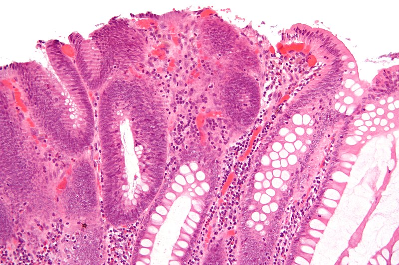
English: High micrograph of a colorectal tubular adenoma without high grade dysplasia. H&E stain.
The lesional tissue, i.e. dysplastic epithelium, is seen on the left of the image and characterized by:
Normal colonic type epithelium is seen on the right of the image and characterized by small round nuclei and abundant goblet cells. The subtle vertical lines (most notable overlying the neoplastic tissue) are an artifact of the tissue preparation. Related imagesThe same case:
Another case:
|
| Source | Own work |
| Author | Nephron |


Licensing
I, the copyright holder of this work, hereby publish it under the following licenses:
This file is licensed under the Creative Commons Attribution-Share Alike 3.0 Unported license.
- You are free:
- to share – to copy, distribute and transmit the work
- to remix – to adapt the work
- Under the following conditions:
- attribution – You must give appropriate credit, provide a link to the license, and indicate if changes were made. You may do so in any reasonable manner, but not in any way that suggests the licensor endorses you or your use.
- share alike – If you remix, transform, or build upon the material, you must distribute your contributions under the same or compatible license as the original.

|
Permission is granted to copy, distribute and/or modify this document under the terms of the GNU Free Documentation License, Version 1.2 or any later version published by the Free Software Foundation; with no Invariant Sections, no Front-Cover Texts, and no Back-Cover Texts. A copy of the license is included in the section entitled GNU Free Documentation License. |
You may select the license of your choice.
| Annotations | This image is annotated: View the annotations at Commons |
File history
Click on a date/time to view the file as it appeared at that time.
| Date/Time | Thumbnail | Dimensions | User | Comment | |
|---|---|---|---|---|---|
| current | 01:07, 27 October 2009 | | 4,272 × 2,848 (3.27 MB) | Nephron | {{Information |Description={{en|1=High micrograph of a colorectal '''tubular adenoma''' without high grade dysplasia. H&E stain. The lesional tissue, i.e. dysplastic epithelium, is seen on the left of the |
File usage
The following page uses this file: