File:Oncocytoma of the Salivary Gland.jpg
Oncocytoma_of_the_Salivary_Gland.jpg (550 × 335 pixels, file size: 25 KB, MIME type: image/jpeg)
Summary
| Description |
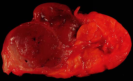
Oncocytoma of the Salivary Gland This lesion presented as a lateral anterior neck mass. At surgery, it was found to be a soft 3.0 x 2.1 x 1.8 cm tumor of the submandibular salivary gland. The photo shows the characteristic dark color of an oncocytoma, a rare type of benign neoplasm, at the left side of the image (the normal lobulated salivary gland tissue is to the right). Excision is curative. Since this specimen was photographed in the fresh state, it is various shades of red due to blood staining. A little formalin fixation would be expected to better emphasize the color difference between the tumor and the normal gland tissue. The photo was shot with a Minolta X-370 with 100mm Rokkor bellows lens on Kodak Elite ISO 100 daylight-balanced transparency film. I used a blue filter to compensate for the tungsten illumination. Photograph by Ed Uthman, MD. Public domain. Posted 19 May 00 |
| Source | http://web2.airmail.net/uthman/specimens/index.html |
| Author | |
| Permission (Reusing this file) |
PD |
Licensing
| This work has been released into the public domain by its author, Ed Uthman. This applies worldwide. In some countries this may not be legally possible; if so: Ed Uthman grants anyone the right to use this work for any purpose, without any conditions, unless such conditions are required by law.
|
File history
Click on a date/time to view the file as it appeared at that time.
| Date/Time | Thumbnail | Dimensions | User | Comment | |
|---|---|---|---|---|---|
| current | 09:53, 5 June 2006 | | 550 × 335 (25 KB) | Patho | {{Information| |Description=Oncocytoma of the Salivary Gland This lesion presented as a lateral anterior neck mass. At surgery, it was found to be a soft 3.0 x 2.1 x 1.8 cm tumor of the submandibular salivary gland. The photo shows the characteristic dar |
File usage
The following page uses this file: