File:Myxomatous aortic valve.jpg
Jump to navigation
Jump to search


Size of this preview: 800 × 600 pixels. Other resolution: 2,048 × 1,536 pixels.
Original file (2,048 × 1,536 pixels, file size: 342 KB, MIME type: image/jpeg)
Summary
| Description |
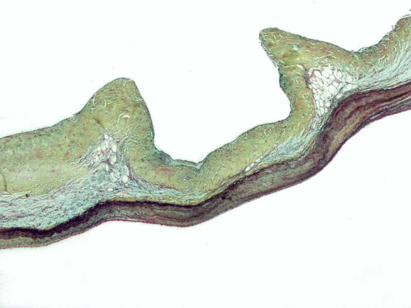
English: Micrograph of myxomatous degeneration of the aortic valve. Surgical specimen. Movat's stain (Black = nuclei, elastic fibres. Yellow = collagen, reticular fibers. Blue =
ground substance, mucin. Bright red = Fibrin. Red = muscle.) In myxomatous degeneration, the ventricularis layer (composed primarily of elastic tissue) is thinned and the spongiosa layer (composed of loose connective tissue) is thickened. On the image, the fibrosa layer (composed of collagen) is on the top, the thickened spongiosa layer below it and the ventricularis layer (made of elastic tissue) at the bottom. The ventricularis layer, as the name may suggest, is closest to the (left) ventricle. The fibrosa layer is closest to the sinus of valsalva. See also
|
| Date | |
| Source | Own work |
| Author | Nephron |
Licensing
I, the copyright holder of this work, hereby publish it under the following licenses:
This file is licensed under the Creative Commons Attribution-Share Alike 3.0 Unported license.
- You are free:
- to share – to copy, distribute and transmit the work
- to remix – to adapt the work
- Under the following conditions:
- attribution – You must give appropriate credit, provide a link to the license, and indicate if changes were made. You may do so in any reasonable manner, but not in any way that suggests the licensor endorses you or your use.
- share alike – If you remix, transform, or build upon the material, you must distribute your contributions under the same or compatible license as the original.

|
Permission is granted to copy, distribute and/or modify this document under the terms of the GNU Free Documentation License, Version 1.2 or any later version published by the Free Software Foundation; with no Invariant Sections, no Front-Cover Texts, and no Back-Cover Texts. A copy of the license is included in the section entitled GNU Free Documentation License. |
You may select the license of your choice.
| Annotations | This image is annotated: View the annotations at Commons |
File history
Click on a date/time to view the file as it appeared at that time.
| Date/Time | Thumbnail | Dimensions | User | Comment | |
|---|---|---|---|---|---|
| current | 05:37, 13 February 2009 | | 2,048 × 1,536 (342 KB) | Nephron | {{Information |Description={{en|1=Myxomatous degeneration of the aortic valve. Surgical specimen. Movat's stain (Black = nuclei, elastic fibres. Yellow = collagen, reticular fibers. Blue = ground substance, mucin. Bright red = Fibrin. Red = muscle.) In m |
File usage
The following page uses this file: