File:Craniopharyngioma1.jpg

From Libre Pathology
Jump to navigation Jump to search

Original file(988 × 1,133 pixels, file size: 177 KB, MIME type: image/jpeg)

This file is from Wikimedia Commons and may be used by other projects. The description on its file description page there is shown below.

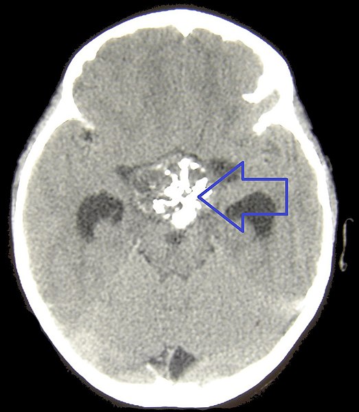
Description CT of a craniopharyngioma. This unenhanced CT shows a calcified cystic structure in the supra sellar region, together with hydrocephalus
Date 6 August 2007 (upload date)
Source Matthew R Garnett, Stéphanie Puget, Jacques Grill, Christian Sainte-Rose. Craniopharyngioma. Orphanet Journal of Rare Diseases. 2, 18. 2007. doi:10.1186/1750-1172-2-18. PMID 17425791
Author see above
w:en:Creative Commons
attribution
This file is licensed under the Creative Commons Attribution 2.0 Generic license.
You are free:
  • to share – to copy, distribute and transmit the work
  • to remix – to adapt the work
Under the following conditions:
  • attribution – You must give appropriate credit, provide a link to the license, and indicate if changes were made. You may do so in any reasonable manner, but not in any way that suggests the licensor endorses you or your use.

Captions

CT of a craniopharyngioma. This unenhanced CT shows a calcified cystic structure in the supra sellar region, together with hydrocephalus.

Elementi ritratti in questo file

depicts

6 August 2007

image/jpeg

File history

Click on a date/time to view the file as it appeared at that time.

Date/TimeThumbnailDimensionsUserComment
current13:55, 11 May 2021Thumbnail for version as of 13:55, 11 May 2021988 × 1,133 (177 KB)Doc JamesArrow

The following page uses this file:

Metadata