File:Choriocarcinoma - very high mag.jpg

Original file (4,272 × 2,848 pixels, file size: 3.74 MB, MIME type: image/jpeg)
Summary
| Description |
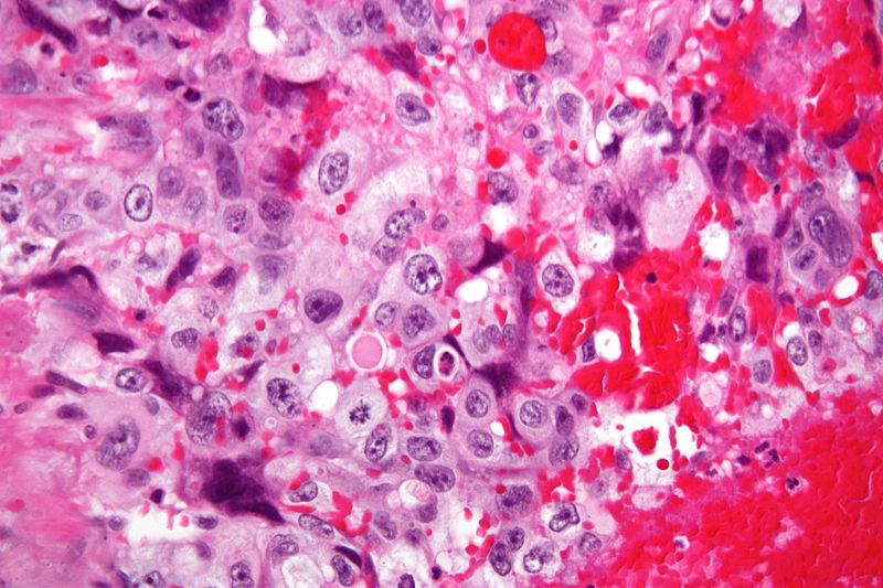
English: Very high magnification micrograph of choriocarcinoma. H&E stain.
Choriocarcinomas consist of two cell populations:
Traditionally, the syncytiotrophoblasts are said to produce the beta-hCG;[1] however, it has been determined that cytotrophoblast also produce some.[2] The syncytiotrophoblasts are often arranged around the outside of cytotrophoblast cell clusters, reminicent of the arrangement in the placenta. On placental villi, the syncytiotrophoblasts are superficial to and, early in pregnancy, cover the cytotrophoblast. Choriocarcinoma is classified as a germ cell tumour. It can arise in the testis or ovary and from a hydatidiform mole. It may be part of a mixed germ cell tumour. Related images
References
|
||
| Source | Own work | ||
| Author | Nephron | ||
| Permission (Reusing this file) |
I, the copyright holder of this work, hereby publish it under the following licenses: This file is licensed under the Creative Commons Attribution-Share Alike 3.0 Unported license.
You may select the license of your choice. |


File history
Click on a date/time to view the file as it appeared at that time.
| Date/Time | Thumbnail | Dimensions | User | Comment | |
|---|---|---|---|---|---|
| current | 04:00, 15 December 2011 | | 4,272 × 2,848 (3.74 MB) | Nephron | {{Information |Description ={{en|1=Very high magnification micrograph of '''choriocarcinoma'''. H&E stain. Choriocarcinomas consist of two cell populations: #Cytotrophoblasts |
File usage
The following 2 pages use this file: