File:Carotid body tumour 2 high mag.jpg

From Libre Pathology
Jump to navigation Jump to search

Original file(4,272 × 2,848 pixels, file size: 6.13 MB, MIME type: image/jpeg)

This file is from Wikimedia Commons and may be used by other projects. The description on its file description page there is shown below.

Summary

Description
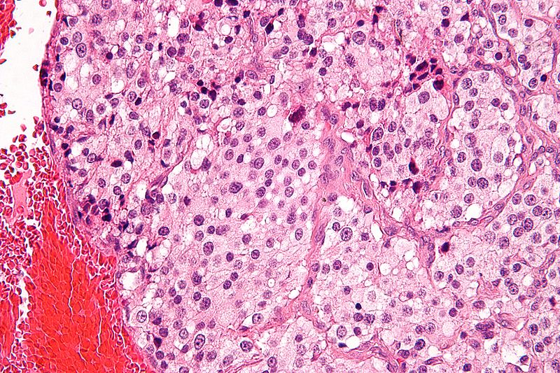
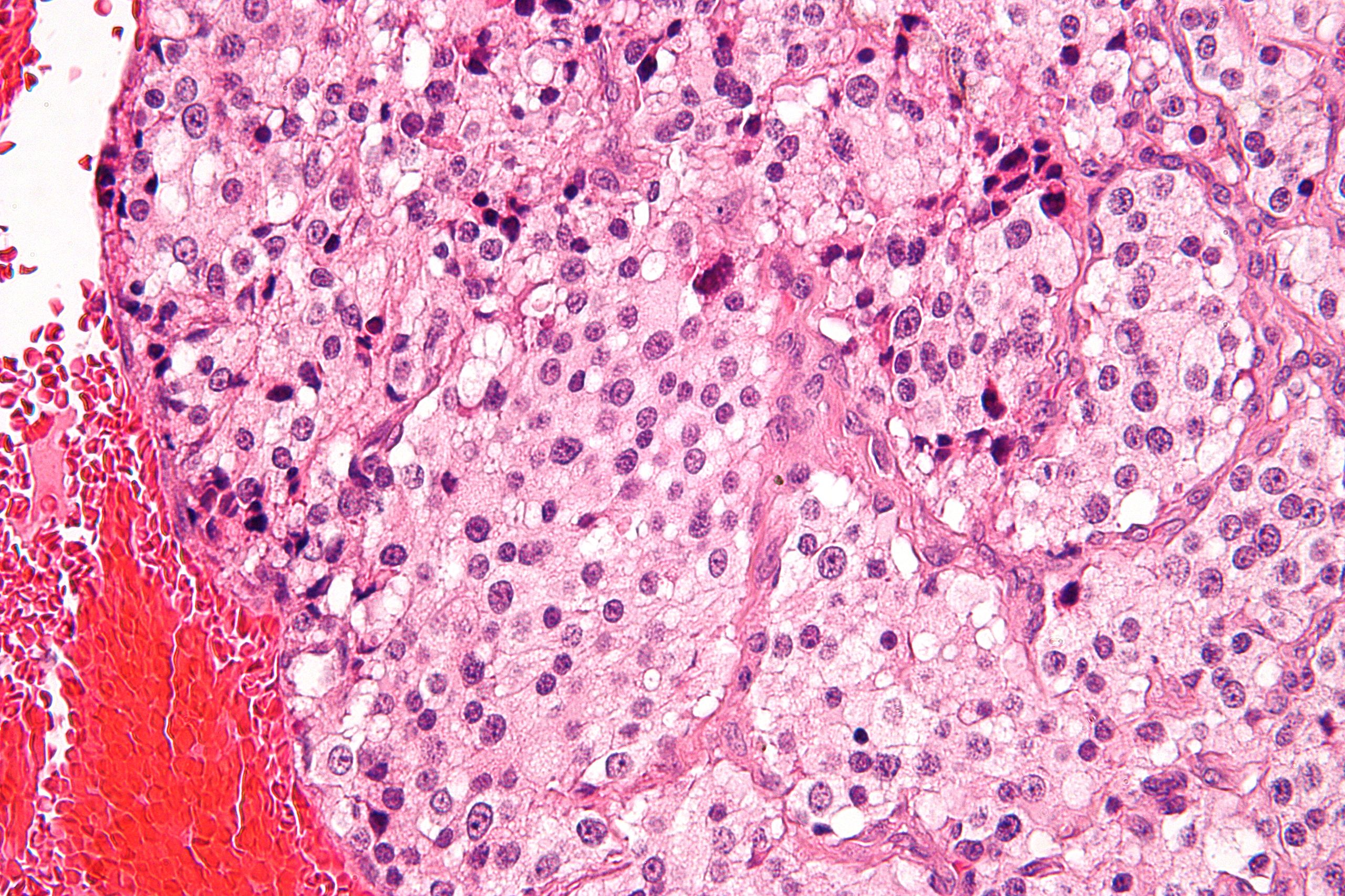
English: High magnification micrograph of a carotid body tumour, a paraganglioma. H&E stain.

Pheochromocytoma is the most common type of paraganglioma.

Histomorphologic features of paragangliomas:

  • Zellballen ("cell balls" in German) - nests of cells.
  • Fibrovascular septae.
  • Finely granular cytoplasm (salt-and-pepper chromatin).

Related images

Source Own work
Author Nephron
Permission
(Reusing this file)
I, the copyright holder of this work, hereby publish it under the following licenses:
w:en:Creative Commons
attribution share alike
This file is licensed under the Creative Commons Attribution-Share Alike 3.0 Unported license.
You are free:
  • to share – to copy, distribute and transmit the work
  • to remix – to adapt the work
Under the following conditions:
  • attribution – You must give appropriate credit, provide a link to the license, and indicate if changes were made. You may do so in any reasonable manner, but not in any way that suggests the licensor endorses you or your use.
  • share alike – If you remix, transform, or build upon the material, you must distribute your contributions under the same or compatible license as the original.
GNU head Permission is granted to copy, distribute and/or modify this document under the terms of the GNU Free Documentation License, Version 1.2 or any later version published by the Free Software Foundation; with no Invariant Sections, no Front-Cover Texts, and no Back-Cover Texts. A copy of the license is included in the section entitled GNU Free Documentation License.
You may select the license of your choice.

Captions

Add a one-line explanation of what this file represents

Items portrayed in this file

depicts

0.03333333333333333333 second

image/jpeg

a7a976e87ec42cea5b801bb26197e54b7df2c782

6,427,587 byte

2,848 pixel

4,272 pixel

File history

Click on a date/time to view the file as it appeared at that time.

Date/TimeThumbnailDimensionsUserComment
current17:45, 17 July 2010Thumbnail for version as of 17:45, 17 July 20104,272 × 2,848 (6.13 MB)Nephron{{Information |Description={{en|1=High magnification micrograph of a '''carotid body tumour''', a paraganglioma. H&E stain. Pheochromocytoma is the most common

The following 2 pages use this file:

Metadata