Wilson's disease
Wilson disease's is a rare autosomal recessive genetic disease characterized by abnormal copper transportation. Its' presentation may be atypical. In the context of pathology, it is typically seen as a liver biopsy.
General
Epidemiology:
- Rare autosomal recessive - mutation in copper-transporting adenosine triphosphatase (ATPase) gene (ATP7B).[1][2]
- Heterozygote carrier rate approximately 1/100 persons.[1]
- Young individuals - usually 12-23 years old.
- May have late-onset (symptomatic when >40 years old).[3]
Clinical:
- Kayser-Fleischer rings --> on slit-lamp examination (green eyes).
- May present to psychiatry or appear to be abusing EtOH.
- Serum ceruloplasmin - lower than normal; typical value for Wilson's ~ 0.12 g/L.
- <0.20 g/L is a criteria for Wilson's disease.[4]
Etiology:
- Excess copper -- due to genetic defect.
Microscopic
Features:
- Nothing specific - known as the great mimicker of liver pathology.
- Steatosis.
- Portal fibrosis.
A. 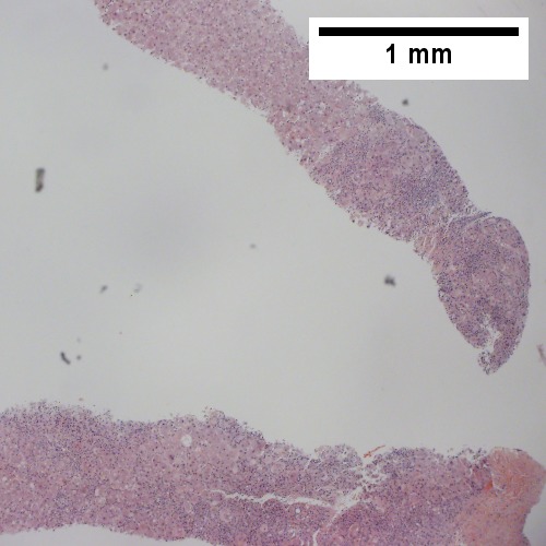
B. ![Bile ductular proliferation with interface hepatitis [inflammation of periportal hepatocytes] (200X).](/w/images/thumb/4/41/2_Wilson_1_680x512px.tif/lossy-page1-500px-2_Wilson_1_680x512px.tif.jpg)
C. 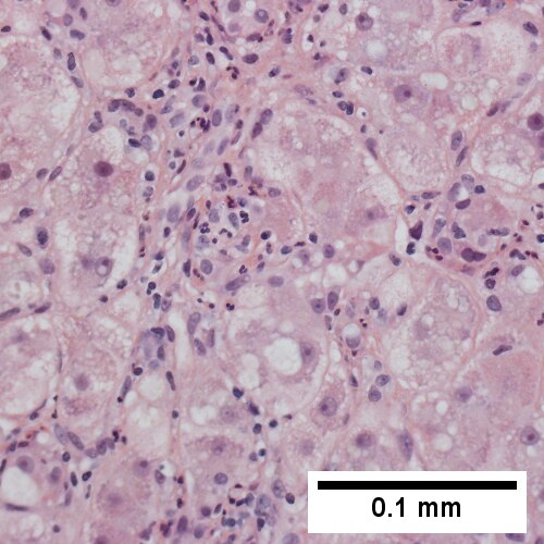
D. 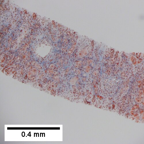
Wilson’s disease, pre-cirrhotic. A. Inflamed & relatively unaffected segments. B. Bile ductular proliferation with interface hepatitis [inflammation of periportal hepatocytes]. C. Enlarged hepatocytes, some with feathery degeneration, others with steatosis. Nuclei show nucleoli. D. Trichrome shows periportal & sinusoidal fibrosis.


Wilson’s disease with cirrhosis in a 22 year old woman. A. Nodules show steatosis without definite triads. B. Trichrome shows minute fibrous bound nodules in this case. C. Reticulin shows extensive regeneration (2-3 nuclei thick cords with lack of direction) with nodule formation. D. Bridge with extensive proliferated bile ductules and acute and chronic inflammatory cells. E. Ballooning degeneration with Mallory bodies.
Stains
- Copper staining positive - only 15% of cases.
- Other stains: rhodinine (red/orange granules = positive), orecin.
Notes:
- Copper staining is a non-specific finding seen in many liver diseases; it is associated with impaired bile secretion.[5]
Image
Additional testing
Copper quantification:
- Mass spectrometry.[6]
- Atomic absorption spectroscopy.
- >250 microg of copper/g of liver suggest Wilson's disease; below does not exclude it.[7]
- 250 microg of copper/g of liver - 80-86% (value dependent on sampling).
- 50 microg of copper/g of liver - 97%.
See also
References
- ↑ 1.0 1.1 http://emedicine.medscape.com/article/183456-overview
- ↑ Online 'Mendelian Inheritance in Man' (OMIM) 606882
- ↑ Ferenci, P.; Członkowska, A.; Merle, U.; Ferenc, S.; Gromadzka, G.; Yurdaydin, C.; Vogel, W.; Bruha, R. et al. (Apr 2007). "Late-onset Wilson's disease.". Gastroenterology 132 (4): 1294-8. doi:10.1053/j.gastro.2007.02.057. PMID 17433323.
- ↑ Diagnostic accuracy of serum ceruloplasmin in Wilson disease: determination of sensitivity and specificity by ROC curve analysis among ATP7B-genotyped subjects. Mak CM, Lam CW, Tam S. Clin Chem. 2008 Aug;54(8):1356-62. Epub 2008 Jun 12. PMID 18556333. URL: http://www.clinchem.org/cgi/reprint/54/8/1356.pdf. Accessed on: 28 September 2009.
- ↑ Miyamura H, Nakanuma Y, Kono N (December 1988). "Survey of copper granules in liver biopsy specimens from various liver abnormalities other than Wilson's disease and biliary diseases". Gastroenterol. Jpn. 23 (6): 633–8. PMID 2464523.
- ↑ Hahn, SH. (May 2014). "Population screening for Wilson's disease.". Ann N Y Acad Sci 1315: 64-9. doi:10.1111/nyas.12423. PMID 24731025.
- ↑ Ferenci, P.; Steindl-Munda, P.; Vogel, W.; Jessner, W.; Gschwantler, M.; Stauber, R.; Datz, C.; Hackl, F. et al. (Aug 2005). "Diagnostic value of quantitative hepatic copper determination in patients with Wilson's Disease.". Clin Gastroenterol Hepatol 3 (8): 811-8. PMID 16234011.
- ↑ Liggi, M.; Mais, C.; Demurtas, M.; Sorbello, O.; Demelia, E.; Civolani, A.; Demelia, L. (Dec 2013). "Uneven distribution of hepatic copper concentration and diagnostic value of double-sample biopsy in Wilson's disease.". Scand J Gastroenterol 48 (12): 1452-8. doi:10.3109/00365521.2013.845904. PMID 24164422.