Venous lake
Jump to navigation
Jump to search
The printable version is no longer supported and may have rendering errors. Please update your browser bookmarks and please use the default browser print function instead.
| Venous lake | |
|---|---|
| Diagnosis in short | |
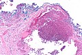
 Venous lake. H&E stain. (WC) | |
| LM DDx | angiokeratoma, cherry hemangioma |
| Gross | classical location: lip |
| Signs | dark blue/purple spot, blanches with pressure |
| Prevalence | common |
| Prognosis | benign |
Venous lake is a benign skin lesion.
General
- Dilated vein.
Clinical:
- Blanches with pressure.[1]
Gross
- Purple/blue spot.
Microscopic
Features:[2]
- Lined by endothelium.
- Blood in lumen.
- +/-Fibrin in lumen.
- +/-Solar elastosis - very common.[3]
DDx:
- Angiokeratoma.
- Ectatic superficial dermal vessels.
- Irregular acanthosis.
- Longer rete ridges.
- Cherry hemangioma.[3]
Images

VL - very low mag. (WC)
VL - low mag. (WC)

Sign out
Left Lower Lip Lesion, Excision:
     - Benign subcutaneous tissue with venous lake.
     - NEGATIVE for malignancy.
Block letters
SKIN LESION, RIGHT CHEEK, BIOPSY:
- VENOUS LAKE.
- SOLAR ELASTOSIS.
- NEGATIVE FOR NEVUS.
Micro
The sections show subcutaneous tissue with dilated vascular spaces. No significant atypia is seen. No overlying skin is present.
See also
References
- ↑ URL: http://dermatlas.med.jhmi.edu/derm/IndexDisplay.cfm?ImageID=-969536424. Accessed on: 13 August 2012.
- ↑ Weedon's Skin Pathology. 3rd Ed. P.895.
- ↑ 3.0 3.1 Busam, Klaus J. (2009). Dermatopathology: A Volume in the Foundations in Diagnostic Pathology Series (1st ed.). Saunders. pp. 551. ISBN 978-0443066542.