Vascular malformations
Jump to navigation
Jump to search
The printable version is no longer supported and may have rendering errors. Please update your browser bookmarks and please use the default browser print function instead.
Vascular malformations come in different flavours.
Types:[1]
- Arteriovenous malformation.
- Most important clinically - highest risk of bleeding.
- Varix.
- One large (dilated) vein.
- Venous angioma.
- Many small veins.
- Cavernous hemangioma (Cavernoma).
- Vessels are back-to-back (no intervening parenchyma).
- Capillary teleangiectasia
- Small capillaries
Also see: Sturge-Weber syndrome.
Arteriovenous malformation
- Abbreviated AVM.
General
- High risk for bleeding vis-a-vis other vascular malformations.
- May be seen in the context of hereditary hemorrhagic telangiectasia.[2]
Gross
Features:[1]
- Classically wedge-shaped - with base toward superficial aspect and apex toward deep aspect (like pulmonary infarcts).
- Usually middle cerebral artery distribution.
Image:
Clinical DDx:
- Melanocytic lesion.
- Other vascular lesions.
Microscopic
Features:
- Large vessels with eccentric wall thickening.
- "Large" = ~ 0.5 mm (0.25-1.0 mm).
- 0.25 mm = ~ 31 RBC diameters across.
- "Large" = ~ 0.5 mm (0.25-1.0 mm).
Notes:
- There is usually one feeding artery.
- The artery is often not seen.
- Arteries have a well-defined internal elastic lamina and an external elastic lamina (best seen on elastic trichrome).
- Veins do not have an external elastic lamina and have a poorly developed/thin internal elastic lamina.
DDx:
- Angioleiomyoma.
- Other vascular lesions.
Images
Cerebral AVM. (WC)

www:
Sign out
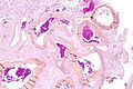
ANUS, BIOPSY:
- ARTERIOVENOUS MALFORMATION (PROMINENT SUPERFICIAL DERMAL ARTERIES AND VEINS).
- SKIN WITH PARAKERATOSIS.
- NEGATIVE FOR MELANOCYTIC LESION.
- NEGATIVE FOR MALIGNANCY.
ANUS, BIOPSY:
- SKIN WITH PARAKERATOSIS.
- PROMINENT SUPERFICIAL DERMAL ARTERIES AND VEINS -- COMPATIBLE WITH
  ARTERIOVENOUS MALFORMATION.
- NEGATIVE FOR MELANOCYTIC LESION.
- NEGATIVE FOR MALIGNANCY.
Cavernous hemangioma
General
- Usually diagnosed by radiology.
- Clinicians call it cavernoma.
Microscopic
Features:
- Vessels back-to-back/little intervening parenchyma.
- Muscle is absent in the vessel walls - key feature.[3]
Images:
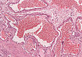
Cavernous hemangioma (WC/Patho82).
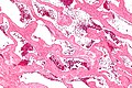
Cavernous hemangioma of the liver (WC).


Venous angioma
General
- Subcortical white matter.
- Most common vascular malformation found at autopsy.
Gross
- May look like petechial hemorrhages.
Microscopy
- Thin walled, dilated vascular channels.
- Lesions larger than capillary teleangiectasia.
Cherry angioma
General
- Benign.
- Common in the elderly.
Clincal:
- Red spot.
- Polypoid.
Gross
- Red spot - well demarcated.
Image:
Microscopic
Features:[5]
- Superficial polypoid lesion that is well-circumscribed.
- Abundant capillaries - key feature.
DDx:
- Venous lake - single vessel.[6]
- Hemangioma.
Images:
Capillary teleangiectasia
- Usually incidental finding at autopsy.
- Pons as predilection site.
Microscopy
- Many small, thin walled capillaries surrounded by normal brain.
- No hemorrhage.
Images:
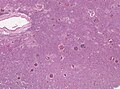
CNS teleangiectasia

See also
References
- ↑ 1.0 1.1 Prayson RA, Kleinschmidt-DeMasters BK (November 2006). "An algorithmic approach to the brain biopsy--part II". Arch. Pathol. Lab. Med. 130 (11): 1639–48. PMID 17076525.
- ↑ Marchuk, DA.; Srinivasan, S.; Squire, TL.; Zawistowski, JS. (Apr 2003). "Vascular morphogenesis: tales of two syndromes.". Hum Mol Genet 12 Spec No 1: R97-112. PMID 12668602.
- ↑ MUN. 23 November 2010.
- ↑ 4.0 4.1 URL:: http://missinglink.ucsf.edu/lm/DermatologyGlossary/cherry_angioma.html. Accessed on: 13 August 2012.
- ↑ Busam, Klaus J. (2009). Dermatopathology: A Volume in the Foundations in Diagnostic Pathology Series (1st ed.). Saunders. pp. 546. ISBN 978-0443066542.
- ↑ Busam, Klaus J. (2009). Dermatopathology: A Volume in the Foundations in Diagnostic Pathology Series (1st ed.). Saunders. pp. 551. ISBN 978-0443066542.