Difference between revisions of "Vascular malformations"
Jump to navigation
Jump to search
(+see also) |
Jensflorian (talk | contribs) (cat) |
||
| (18 intermediate revisions by 2 users not shown) | |||
| Line 8: | Line 8: | ||
#Venous angioma. | #Venous angioma. | ||
#*Many small veins. | #*Many small veins. | ||
# | #Cavernous hemangioma (Cavernoma). | ||
#*Vessels are back-to-back (no intervening parenchyma). | #*Vessels are back-to-back (no intervening parenchyma). | ||
#Capillary teleangiectasia | |||
#*Small capillaries | |||
Also see: ''[[Sturge-Weber syndrome]]''. | Also see: ''[[Sturge-Weber syndrome]]''. | ||
==Arteriovenous malformation== | ==Arteriovenous malformation== | ||
*Abbreviated ''AVM''. | |||
===General=== | ===General=== | ||
*High risk for bleeding vis-a-vis other vascular malformations. | *High risk for bleeding vis-a-vis other vascular malformations. | ||
*May be seen in the context of [[hereditary hemorrhagic telangiectasia]].<ref name=pmid12668602>{{Cite journal | last1 = Marchuk | first1 = DA. | last2 = Srinivasan | first2 = S. | last3 = Squire | first3 = TL. | last4 = Zawistowski | first4 = JS. | title = Vascular morphogenesis: tales of two syndromes. | journal = Hum Mol Genet | volume = 12 Spec No 1 | issue = | pages = R97-112 | month = Apr | year = 2003 | doi = | PMID = 12668602 }}</ref> | |||
===Gross=== | ===Gross=== | ||
| Line 21: | Line 25: | ||
*Classically wedge-shaped - with base toward superficial aspect and apex toward deep aspect (like pulmonary infarcts). | *Classically wedge-shaped - with base toward superficial aspect and apex toward deep aspect (like pulmonary infarcts). | ||
*Usually middle cerebral artery distribution. | *Usually middle cerebral artery distribution. | ||
Image: | |||
*[http://www.flickr.com/photos/11462589@N05/1125883661/in/photostream/ AVM (flickr.com)]. | |||
Clinical DDx: | |||
*Melanocytic lesion. | |||
*Other vascular lesions. | |||
===Microscopic=== | ===Microscopic=== | ||
| Line 27: | Line 38: | ||
**"Large" = ~ 0.5 mm (0.25-1.0 mm). | **"Large" = ~ 0.5 mm (0.25-1.0 mm). | ||
***0.25 mm = ~ 31 RBC diameters across. | ***0.25 mm = ~ 31 RBC diameters across. | ||
Notes: | Notes: | ||
| Line 36: | Line 45: | ||
**[[Vein]]s do not have an external elastic lamina and have a poorly developed/thin internal elastic lamina. | **[[Vein]]s do not have an external elastic lamina and have a poorly developed/thin internal elastic lamina. | ||
==Cavernous | DDx: | ||
*Other vascular lesions. | |||
====Images==== | |||
<gallery> | |||
Image:Arteriovenous_malformation_-_brain_-_low_mag.jpg | Cerebral AVM. (WC) | |||
</gallery> | |||
www: | |||
*[http://path.upmc.edu/cases/case123.html AVM - several images (upmc.edu)]. | |||
===Sign out=== | |||
<pre> | |||
ANUS, BIOPSY: | |||
- ARTERIOVENOUS MALFORMATION (PROMINENT SUPERFICIAL DERMAL ARTERIES AND VEINS). | |||
- SKIN WITH PARAKERATOSIS. | |||
- NEGATIVE FOR MELANOCYTIC LESION. | |||
- NEGATIVE FOR MALIGNANCY. | |||
</pre> | |||
<pre> | |||
ANUS, BIOPSY: | |||
- SKIN WITH PARAKERATOSIS. | |||
- PROMINENT SUPERFICIAL DERMAL ARTERIES AND VEINS -- COMPATIBLE WITH | |||
ARTERIOVENOUS MALFORMATION. | |||
- NEGATIVE FOR MELANOCYTIC LESION. | |||
- NEGATIVE FOR MALIGNANCY. | |||
</pre> | |||
==Cavernous hemangioma== | |||
===General=== | ===General=== | ||
*Usually diagnosed by radiology. | *Usually diagnosed by radiology. | ||
*Clinicians call it cavernoma. | |||
===Microscopic=== | ===Microscopic=== | ||
| Line 44: | Line 83: | ||
*Vessels back-to-back/little intervening parenchyma. | *Vessels back-to-back/little intervening parenchyma. | ||
**Muscle is absent in the vessel walls - '''key feature'''.<ref>MUN. 23 November 2010.</ref> | **Muscle is absent in the vessel walls - '''key feature'''.<ref>MUN. 23 November 2010.</ref> | ||
Images: | |||
<gallery> | |||
File:Cavernous hemangioma.jpg | Cavernous hemangioma (WC/Patho82). | |||
File:Cavernous liver hemangioma - intermed mag.jpg | Cavernous hemangioma of the liver (WC). | |||
</gallery> | |||
==Venous angioma== | |||
===General=== | |||
*Subcortical white matter. | |||
*Most common vascular malformation found at autopsy. | |||
===Gross=== | |||
*May look like petechial hemorrhages. | |||
===Microscopy=== | |||
*Thin walled, dilated vascular channels. | |||
*Lesions larger than capillary teleangiectasia. | |||
==Cherry angioma== | |||
*[[AKA]] ''Campbell De Morgan spots''. | |||
*[[AKA]] ''senile angioma''. | |||
*[[AKA]] ''cherry hemangioma''. | |||
===General=== | |||
*Benign. | |||
*Common in the elderly. | |||
Clincal: | |||
*Red spot. | |||
*Polypoid. | |||
===Gross=== | |||
*Red spot - well demarcated. | |||
Image: | |||
*[http://missinglink.ucsf.edu/lm/DermatologyGlossary/img/Dermatology%20Glossary/Glossary%20Clinical%20Images/CherryAngioma.jpg Cherry angioma (ucsf.edu)].<ref name=ucsf_ca>URL:: [http://missinglink.ucsf.edu/lm/DermatologyGlossary/cherry_angioma.html http://missinglink.ucsf.edu/lm/DermatologyGlossary/cherry_angioma.html]. Accessed on: 13 August 2012.</ref> | |||
===Microscopic=== | |||
Features:<ref>{{Ref Derm|546}}</ref> | |||
*Superficial polypoid lesion that is well-circumscribed. | |||
*Abundant capillaries - '''key feature'''. | |||
DDx: | |||
*[[Venous lake]] - single vessel.<ref name=Ref_Derm551>{{Ref Derm|551}}</ref> | |||
*[[Hemangioma]]. | |||
Images: | |||
*[http://www.dermpathexpert.com/id30.html Cherry hemangioma (dermpathexpert.com)]. | |||
*[http://missinglink.ucsf.edu/lm/DermatologyGlossary/img/Dermatology%20Glossary/Glossary%20Histo%20Images/cherry_angioma_mid_power.jpg Cherry angioma (ucsf.edu)].<ref name=ucsf_ca>URL:: [http://missinglink.ucsf.edu/lm/DermatologyGlossary/cherry_angioma.html http://missinglink.ucsf.edu/lm/DermatologyGlossary/cherry_angioma.html]. Accessed on: 13 August 2012.</ref> | |||
==Capillary teleangiectasia== | |||
*Usually incidental finding at autopsy. | |||
*Pons as predilection site. | |||
===Microscopy=== | |||
*Many small, thin walled capillaries surrounded by normal brain. | |||
*No hemorrhage. | |||
Images: | |||
<gallery> | |||
File:Teleangiectasia brain.jpg | CNS teleangiectasia | |||
</gallery> | |||
==See also== | ==See also== | ||
| Line 49: | Line 154: | ||
*[[Vascular tumours]]. | *[[Vascular tumours]]. | ||
*[[Vasculitis]]. | *[[Vasculitis]]. | ||
*[[Littoral cell angioma]]. | |||
==References== | ==References== | ||
{{Reflist|2}} | {{Reflist|2}} | ||
[[Category:Malformation]] | |||
[[Category:Cardiovascular pathology]] | [[Category:Cardiovascular pathology]] | ||
[[Category:Neuropathology]] | [[Category:Neuropathology]] | ||
Revision as of 13:12, 14 October 2019
Vascular malformations come in different flavours.
Types:[1]
- Arteriovenous malformation.
- Most important clinically - highest risk of bleeding.
- Varix.
- One large (dilated) vein.
- Venous angioma.
- Many small veins.
- Cavernous hemangioma (Cavernoma).
- Vessels are back-to-back (no intervening parenchyma).
- Capillary teleangiectasia
- Small capillaries
Also see: Sturge-Weber syndrome.
Arteriovenous malformation
- Abbreviated AVM.
General
- High risk for bleeding vis-a-vis other vascular malformations.
- May be seen in the context of hereditary hemorrhagic telangiectasia.[2]
Gross
Features:[1]
- Classically wedge-shaped - with base toward superficial aspect and apex toward deep aspect (like pulmonary infarcts).
- Usually middle cerebral artery distribution.
Image:
Clinical DDx:
- Melanocytic lesion.
- Other vascular lesions.
Microscopic
Features:
- Large vessels with eccentric wall thickening.
- "Large" = ~ 0.5 mm (0.25-1.0 mm).
- 0.25 mm = ~ 31 RBC diameters across.
- "Large" = ~ 0.5 mm (0.25-1.0 mm).
Notes:
- There is usually one feeding artery.
- The artery is often not seen.
- Arteries have a well-defined internal elastic lamina and an external elastic lamina (best seen on elastic trichrome).
- Veins do not have an external elastic lamina and have a poorly developed/thin internal elastic lamina.
DDx:
- Other vascular lesions.
Images
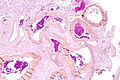
Cerebral AVM. (WC)

www:
Sign out
ANUS, BIOPSY:
- ARTERIOVENOUS MALFORMATION (PROMINENT SUPERFICIAL DERMAL ARTERIES AND VEINS).
- SKIN WITH PARAKERATOSIS.
- NEGATIVE FOR MELANOCYTIC LESION.
- NEGATIVE FOR MALIGNANCY.
ANUS, BIOPSY:
- SKIN WITH PARAKERATOSIS.
- PROMINENT SUPERFICIAL DERMAL ARTERIES AND VEINS -- COMPATIBLE WITH
  ARTERIOVENOUS MALFORMATION.
- NEGATIVE FOR MELANOCYTIC LESION.
- NEGATIVE FOR MALIGNANCY.
Cavernous hemangioma
General
- Usually diagnosed by radiology.
- Clinicians call it cavernoma.
Microscopic
Features:
- Vessels back-to-back/little intervening parenchyma.
- Muscle is absent in the vessel walls - key feature.[3]
Images:
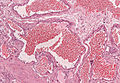
Cavernous hemangioma (WC/Patho82).
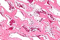
Cavernous hemangioma of the liver (WC).


Venous angioma
General
- Subcortical white matter.
- Most common vascular malformation found at autopsy.
Gross
- May look like petechial hemorrhages.
Microscopy
- Thin walled, dilated vascular channels.
- Lesions larger than capillary teleangiectasia.
Cherry angioma
General
- Benign.
- Common in the elderly.
Clincal:
- Red spot.
- Polypoid.
Gross
- Red spot - well demarcated.
Image:
Microscopic
Features:[5]
- Superficial polypoid lesion that is well-circumscribed.
- Abundant capillaries - key feature.
DDx:
- Venous lake - single vessel.[6]
- Hemangioma.
Images:
Capillary teleangiectasia
- Usually incidental finding at autopsy.
- Pons as predilection site.
Microscopy
- Many small, thin walled capillaries surrounded by normal brain.
- No hemorrhage.
Images:
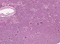
CNS teleangiectasia

See also
References
- ↑ 1.0 1.1 Prayson RA, Kleinschmidt-DeMasters BK (November 2006). "An algorithmic approach to the brain biopsy--part II". Arch. Pathol. Lab. Med. 130 (11): 1639–48. PMID 17076525.
- ↑ Marchuk, DA.; Srinivasan, S.; Squire, TL.; Zawistowski, JS. (Apr 2003). "Vascular morphogenesis: tales of two syndromes.". Hum Mol Genet 12 Spec No 1: R97-112. PMID 12668602.
- ↑ MUN. 23 November 2010.
- ↑ 4.0 4.1 URL:: http://missinglink.ucsf.edu/lm/DermatologyGlossary/cherry_angioma.html. Accessed on: 13 August 2012.
- ↑ Busam, Klaus J. (2009). Dermatopathology: A Volume in the Foundations in Diagnostic Pathology Series (1st ed.). Saunders. pp. 546. ISBN 978-0443066542.
- ↑ Busam, Klaus J. (2009). Dermatopathology: A Volume in the Foundations in Diagnostic Pathology Series (1st ed.). Saunders. pp. 551. ISBN 978-0443066542.