Difference between revisions of "Vascular disease"
(→Aortic dissection: split out) |
|||
| Line 77: | Line 77: | ||
==Aortic dissection== | ==Aortic dissection== | ||
*Abbreviated ''AoD''. | *Abbreviated ''AoD''. | ||
{{Main|Aortic dissection}} | |||
==Cystic medial degeneration== | ==Cystic medial degeneration== | ||
Revision as of 13:06, 13 May 2014
The article covers vascular disease, i.e. diseases of blood vessels. These keep vascular surgeons and cardiac surgeon busy.
Vasculitides are covered in a separate article called vasculitides.
Normal blood vessels
Comparing arteries and veins:[1]
| Feature | Artery | Vein |
|---|---|---|
| Internal elastic lamina (IEL) | prominent/thick, usu. complete | thin & incomplete |
| External elastic lamina (EEL) | present, thick | absent |
| Shape | circular / lumen wide open | collapsed |
| Wall thickness | thick | thin |

Great vessels
When things go wrong here, you see a cardiac surgeon.
Atherosclerosis
General
- A leading cause of death, esp. in the Western world.
- May have multi-system manifestations.
Location and associated pathology:
- Coronary artery atherosclerosis (AKA coronary artery disease) -> myocardial infarction +/-coronary thrombosis.
- Atherosclerotic peripheral vascular disease -> leg amputations.
- Carotid artery atherosclerosis -> thrombotic stroke.
- Superior mesenteric artery atherosclerosis -> ischemic enteritis or ischemic colitis or ischemic enterocolitis.
- Penile artery atherosclerosis -> impotence.
Clinical risk factors:
- Age.
- Blood pressure (high) - modifiable (antihypertensives).
- Cholesterol - modifiable (statins, diet).
- Diabetes mellitus - modifiable (hypoglycemic medications, diet, lifestyle).
- Smoking - modifiable (cessation).
- Family history.
Microscopic
Features:
- Intimal hyperplasia.
- Lipid deposition.
- Foamy macrophages within intima & media.
- Cholesterol clefts
- Luminal narrowing.
Notes:
- Considered "complex" if any of the following are present:[2]
- Calcifications.
- Thrombosis.
- Haemorrhage.
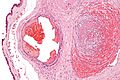
Image
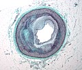
Right coronary artery with atherosclerosis. (WC/Nephron)

Stains
- Elastic trichrome stain or Movat stain - highlights duplication of internal elastic lamina, allows on to identify with ease intimal thickening.
Aortic dissection
- Abbreviated AoD.
Cystic medial degeneration
General
- Nonspecific finding - may be seen in a number of conditions.
Note about cystic medial necrosis:
- Often not cystic and not necrotic.
Microscopic
- Basophilic ground substance in the media (seen on Movat's stain).
- Disruption of the elastic lamina (seen on elastic trichrome stain).
- +/-Focal necrosis.
Images
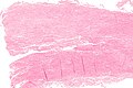
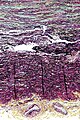
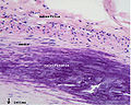
Cystic medial degeneration - low mag. (WC/Nephron)
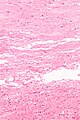
Cystic medial degeneration - high mag. (WC/Nephron)
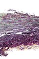
Cystic medial degeneration - movat - low mag. (WC/Nephron)
Cystic medial degeneration - movat - intermed. mag. (WC/Nephron)


www:
Stains
- Elastin stains (e.g. elastic trichrome stain) - disruption of the elastic lamina.
- Movat's stain - basophilic ground substance in the media.
Medial calcific sclerosis
- AKA Moenckeberg medial calcific sclerosis, calcific medial sclerosis of Monckeberg, and Monckeberg's arteriosclerosis.
General
- Usually of no clinical consequence.
Microscopic
Features:[6]
- Medial calcification (purple irregular stuff on H&E -- calcium phosphate).
Note:
- Lumen unaffected.
Images
MCS. (WC)

www:
Sign out
RIGHT LEG, BELOW KNEE AMPUTATION:
- MINIMAL-TO-MILD LARGE VESSEL ATHEROSCLEROSIS, SEE COMMENT.
- MEDIAL CALCIFIC SCLEROSIS.
- SKIN WITH DERMAL FIBROSIS.

COMMENT:
The sections may not be representative of disease in the distal vascular bed.
Hyperplastic arteriolosclerosis
General
- Associated with:[7]
- Malignant hypertension.
- Scleroderma.
- May be a consequence of thrombotic microangiopathy.[citation needed]
Note:
- Hyperplasia = proliferation of cells.
Microscopic
Features:[6]
- Onion-skin appearance of intima & media due to:
- Intimal hyperplasia.
- Smooth muscle hyperplasia.
Image: Hyperplastic arteriolosclerosis (utah.edu).
Fibromuscular dysplasia
- Abbreviated FMD.
General
Etiology:
- Unknown, possibly genetic.
Gender:
- Women > men.
- May be seen in virtually any artery.
- Reported as a cause of sudden death with involvement of the artery supplying the AV node.[8]
Gross/radiologic
- Segmental - thinning and thickening.[9]
Classical locations:[9]
- Renal artery - leading to hypertension.
- Carotid artery.
Microscopic
Features:[9]
- Smooth muscle hyperplasia - key feature.
- Elastic fibre fragmentation.
- Luminal narrowing.
Images:
Stains
- Elastic trichrome or Movat stain - to demonstrate elastic fibre fragmentation.
Thromboangiitis obliterans
Thrombosis
- See also: Cerebral venous thrombosis.
General
Definition:
- Blood clot formation within a vessel.
Complications:
- Embolism - see: Pulmonary thromboembolism.
Risk factors:
- The classic pimping question is what "Virchow's triad?"
- Stasis, hypercoagulability, endothelial injury.
- A long list is found in: risk factors for VTE.
Gross
Microscopic
Features:
- Lines of Zahn.
- Fibrin - pink acellular stuff on a H&E stain.
Image
Intravascular fibrin - high mag. (WC/Nephron)

Cholesterol embolism
- Abbreviated CE.
Coarctation of the aorta
- AKA aortic coarctation.
General
- Uncommon.
Classification:
- Preductal.
- Postductal.
Associations:
Clinical
Presentation:[12]
- Heart failure.
- Hypertension - esp. upper extremity vs. lower extremity.
Gross
- Narrowing (stenosis) of the aorta proximal or distal to the ductus arteriosis.
Image
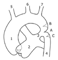
Pre- and postductal coarctation of the aorta - schematic (WC)

Intracranial berry aneurysm
See also
References
- ↑ URL: http://www.lab.anhb.uwa.edu.au/mb140/corepages/vascular/vascular.htm. Accessed on: 13 January 2011.
- ↑ Klatt, Edward C. (2006). Robbins and Cotran Atlas of Pathology (1st ed.). Saunders. pp. 4. ISBN 978-1416002741.
- ↑ URL: http://emedicine.medscape.com/article/756835-overview. Accessed on: 12 August 2010.
- ↑ URL: http://emedicine.medscape.com/article/756835-overview. Accessed on: 12 August 2010.
- ↑ Ha HI, Seo JB, Lee SH, et al. (2007). "Imaging of Marfan syndrome: multisystemic manifestations". Radiographics 27 (4): 989–1004. doi:10.1148/rg.274065171. PMID 17620463. http://radiographics.rsna.org/content/27/4/989.full.
- ↑ 6.0 6.1 Klatt, Edward C. (2006). Robbins and Cotran Atlas of Pathology (1st ed.). Saunders. pp. 7. ISBN 978-1416002741.
- ↑ URL: http://library.med.utah.edu/WebPath/IMMHTML/IMM028.html. Accessed on: 11 May 2011.
- ↑ 8.0 8.1 Lee, S.; Chae, J.; Cho, Y. (Dec 2006). "Causes of sudden death related to sexual activity: results of a medicolegal postmortem study from 2001 to 2005.". J Korean Med Sci 21 (6): 995-9. PMID 17179675.
- ↑ 9.0 9.1 9.2 Hata, D. (Sep 2001). "Fibromuscular dysplasia.". Intern Med 40 (9): 978-9. PMID 11579971.
- ↑ Braverman, AC.; Güven, H.; Beardslee, MA.; Makan, M.; Kates, AM.; Moon, MR. (Sep 2005). "The bicuspid aortic valve.". Curr Probl Cardiol 30 (9): 470-522. doi:10.1016/j.cpcardiol.2005.06.002. PMID 16129122.
- ↑ Hjerrild, BE.; Mortensen, KH.; Sørensen, KE.; Pedersen, EM.; Andersen, NH.; Lundorf, E.; Hansen, KW.; Hørlyck, A. et al. (2010). "Thoracic aortopathy in Turner syndrome and the influence of bicuspid aortic valves and blood pressure: a CMR study.". J Cardiovasc Magn Reson 12: 12. doi:10.1186/1532-429X-12-12. PMID 20222980.
- ↑ Peres, A.; Martins, JD.; Paramés, F.; Gil, R.; Matias, C.; Franco, J.; Freitas, I.; Trigo, C. et al. (Jan 2010). "Isolated aortic coarctation: experience in 100 consecutive patients.". Rev Port Cardiol 29 (1): 23-35. PMID 20391897.