Difference between revisions of "User:Michael/Case"
Jump to navigation
Jump to search
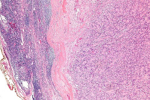
Low magnification. H&E stain.
m (Michael moved page User:Michael/Snapper to User:Michael/Case) |
|||
| (One intermediate revision by one other user not shown) | |||
| Line 6: | Line 6: | ||
<center>Low magnification. [[H&E stain]].</center> | <center>Low magnification. [[H&E stain]].</center> | ||
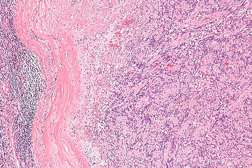
{{hidden| | {{hidden|Intermediate magnification|[[Image:Intranodal palisaded myofibroblastoma - intermed mag.jpg|500px|link=|center|]] | ||
<center>Intermediate magnification. [[H&E stain]].</center>}} | <center>Intermediate magnification. [[H&E stain]].</center>}} | ||
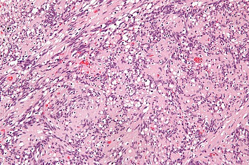
{{hidden| | {{hidden|High magnification|[[Image:Intranodal palisaded myofibroblastoma - high mag.jpg|500px|link=|center|]] | ||
<center>High magnification. [[H&E stain]].</center>}} | <center>High magnification. [[H&E stain]].</center>}} | ||
Latest revision as of 18:31, 14 January 2014
Clinical history
65 year old man, enlarged inguinal lymph node.
Morphology
Intermediate magnification
|
|---|
 |
High magnification
|
|---|
 |
Differential diagnosis
Differential diagnosis
|
|---|
|
|
Additional tests
Additional tests
|
|---|
|
|
Additional tests
|
|---|
|
|
Diagnosis
Diagnosis
|
|---|
|
|