Difference between revisions of "Urachal carcinoma"
Jump to navigation
Jump to search
(split-out) |
(+infobox) |
||
| Line 1: | Line 1: | ||
{{ Infobox diagnosis | |||
| Name = {{PAGENAME}} | |||
| Image = Urachal_carcinoma_-_high_mag.jpg | |||
| Width = | |||
| Caption = Urachal carcinoma. [[H&E stain]]. | |||
| Micro = atypical cells - usually gland forming, +/-mucinous, +/-signet rings | |||
| Subtypes = enteric, mucinous, signet ring | |||
| LMDDx = [[adenocarcinoma of the urinary bladder]], invasive [[urothelial carcinoma]] with glandular differentiation, [[metastatic]] adenocarcinoma | |||
| Stains = | |||
| IHC = CK20 +ve, beta-catenin +ve (non-nuclear), p63 -ve, CK34betaE12 +ve | |||
| EM = | |||
| Molecular = | |||
| IF = | |||
| Gross = | |||
| Grossing = | |||
| Site = [[urinary bladder]] - specifically the dome | |||
| Assdx = | |||
| Syndromes = | |||
| Clinicalhx = | |||
| Signs = +/-hematuria | |||
| Symptoms = | |||
| Prevalence = very rare | |||
| Bloodwork = | |||
| Rads = | |||
| Endoscopy = | |||
| Prognosis = usually poor | |||
| Other = | |||
| ClinDDx = other bladder tumours - esp. [[urothelial carcinoma]] | |||
}} | |||
'''Urachal carcinoma''' is an uncommon [[cancer|malignant tumour]] that arises from the urachus. Most urachal carcinomas are ''[[adenocarcinoma]]s''. | '''Urachal carcinoma''' is an uncommon [[cancer|malignant tumour]] that arises from the urachus. Most urachal carcinomas are ''[[adenocarcinoma]]s''. | ||
Revision as of 00:23, 6 December 2013
| Urachal carcinoma | |
|---|---|
| Diagnosis in short | |
 Urachal carcinoma. H&E stain. | |
|
| |
| LM | atypical cells - usually gland forming, +/-mucinous, +/-signet rings |
| Subtypes | enteric, mucinous, signet ring |
| LM DDx | adenocarcinoma of the urinary bladder, invasive urothelial carcinoma with glandular differentiation, metastatic adenocarcinoma |
| IHC | CK20 +ve, beta-catenin +ve (non-nuclear), p63 -ve, CK34betaE12 +ve |
| Site | urinary bladder - specifically the dome |
|
| |
| Signs | +/-hematuria |
| Prevalence | very rare |
| Prognosis | usually poor |
| Clin. DDx | other bladder tumours - esp. urothelial carcinoma |
Urachal carcinoma is an uncommon malignant tumour that arises from the urachus. Most urachal carcinomas are adenocarcinomas.
General
Treatment:
- Partial cystectomy +/- umbilectomy.
Gross
- Lesion must be in urachus or dome of urinary bladder.
Microscopic
Features:
- Usually adenocarcinoma.
- Adjacent urothelium typically benign.
DDx:[3]
- Adenocarcinoma of the urinary bladder.
- Invasive urothelial carcinoma with glandular differentiation.
- Metastatic adenocarcinoma/adenocarcinoma extending from another structure, e.g. colorectal adenocarcinoma.
Patterns
- Enteric - looks like colonic adenocarcinoma.
- Mucinous.
- Signet ring.
Note:
- Urachal carcinoma may be nonglandular.[4]
Images
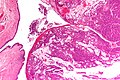
UC - very low mag. (WC)
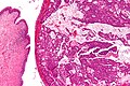
UC - low mag. (WC)
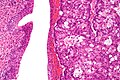
UC - high mag. (WC)


IHC
Features:[5]
- CK20 +ve.
- CK7 +ve/-ve.
- CK34betaE12 +ve/-ve.
- Beta-catenin -- usu cytoplasmic/membranous +ve.
Others:[6]
- p63 -ve (+ve in only 3%).
UC versus CRC -- not absolute but useful:
- CK34betaE12 +ve in UC (-ve in CRC).
- Beta-catenin -ve nuclei in UC (+ve nuclei in CRC).
Sign out
- The diagnosis is clinicopathologic; one needs imaging.[3]
See also
References
- ↑ Ashley, RA.; Inman, BA.; Sebo, TJ.; Leibovich, BC.; Blute, ML.; Kwon, ED.; Zincke, H. (Aug 2006). "Urachal carcinoma: clinicopathologic features and long-term outcomes of an aggressive malignancy.". Cancer 107 (4): 712-20. doi:10.1002/cncr.22060. PMID 16826585.
- ↑ Bruins, HM.; Visser, O.; Ploeg, M.; Hulsbergen-van de Kaa, CA.; Kiemeney, LA.; Witjes, JA. (Oct 2012). "The clinical epidemiology of urachal carcinoma: results of a large, population based study.". J Urol 188 (4): 1102-7. doi:10.1016/j.juro.2012.06.020. PMID 22901574.
- ↑ 3.0 3.1 Amin, Mahul B. (2010). Diagnostic Pathology: Genitourinary (1st ed.). Amirsys. pp. 2-143. ISBN 978-1931884280.
- ↑ Paner, GP.; Barkan, GA.; Mehta, V.; Sirintrapun, SJ.; Tsuzuki, T.; Sebo, TJ.; Jimenez, RE. (Mar 2012). "Urachal carcinomas of the nonglandular type: salient features and considerations in pathologic diagnosis.". Am J Surg Pathol 36 (3): 432-42. doi:10.1097/PAS.0b013e31823fe49c. PMID 22301493.
- ↑ Gopalan, A.; Sharp, DS.; Fine, SW.; Tickoo, SK.; Herr, HW.; Reuter, VE.; Olgac, S. (May 2009). "Urachal carcinoma: a clinicopathologic analysis of 24 cases with outcome correlation.". Am J Surg Pathol 33 (5): 659-68. doi:10.1097/PAS.0b013e31819aa4ae. PMID 19252435.
- ↑ Paner, GP.; McKenney, JK.; Barkan, GA.; Yao, JL.; Frankel, WL.; Sebo, TJ.; Shen, SS.; Jimenez, RE. (Jun 2011). "Immunohistochemical analysis in a morphologic spectrum of urachal epithelial neoplasms: diagnostic implications and pitfalls.". Am J Surg Pathol 35 (6): 787-98. doi:10.1097/PAS.0b013e3182189c11. PMID 21572312.