Synovial sarcoma
Jump to navigation
Jump to search
The printable version is no longer supported and may have rendering errors. Please update your browser bookmarks and please use the default browser print function instead.
| Synovial sarcoma | |
|---|---|
| Diagnosis in short | |
 Monophasic synovial sarcoma. H&E stain. | |
|
| |
| LM | one of the following: (1) spindle cell sarcoma with features of hemangiopericytoma, i.e. staghorn vessels; (2) biphasic synovial sarcoma (spindle cells with features of hemangiopericytoma, epitheliod glands or nests); (3) primitive round cell type |
| LM DDx | MPNST, hemangiopericytoma, fibrosarcoma, small round cell tumours, carcinoma |
| IHC | Vimentin +ve, EMA +ve, BCL2 +ve, CD99 +ve. |
| Molecular | t(X;18) |
| Gross | usually lower extremity, usually close to a joint |
| Site | soft tissue |
|
| |
| Clinical history | young adults or adolescents |
| Signs | mass +/-pain |
| Prevalence | uncommon |
| Prognosis | poor |
Synovial sarcoma, abbreviated SS, is an uncommon malignant soft tissue tumour, typically seen in young adults.
General
- Does not arise from cartilage.[1]
- Young adults or adolescents.
- Classic age: 30s.
- Poor prognosis.
Clinical:[2]
- Present with soft palpable mass - slow growing - often for years.
- May present with pain.
- Uncommon finding in sarcomas.
Gross
Location:
- Usually close to a joint.
- Usually distal extremity ~65% of cases.[2]
- Upper extremity ~20% of cases.[2]
Appearance - often non-specific:
- Solid often lobulated +/- cystic component.
- Grey-yellow.
- Pushing border to ill-defined border.
Images:
Microscopic
Comes in three (histologic) flavours:[1][4]
- Spindle cell sarcoma with features of hemangiopericytoma, i.e. staghorn vessels.
- Biphasic synovial sarcoma:
- Spindle cells with features of hemangiopericytoma.
- Epitheliod glands or nests.
- Primitive round cell type.
Features:
- Herring bone or vesicular pattern - key feature.
- Spindle cells.
- +/-Glandular component - typically more pink.
- +/-Calcification - uncommon.
- Extensive calcification = better prognosis.[5]
DDx:
- MPNST.
- Can be difficult.
Images
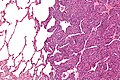
Monophasic synovial sarcoma with staghorn vessels - intermed. mag. (WC/Nephron)
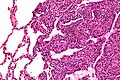
Monophasic synovial sarcoma with staghorn vessels - high mag. (WC/Nephron)

www:
- Synovial sarcoma (scielo.br).
- Synovial sarcoma - collection of images (humpath.com).
- Synovial sarcoma - several images (upmc.edu).
- Biphasic SS (radiographics.rsna.org).
- Monophasic SS (radiographics.rsna.org).
IHC
Features:[1]
- Vimentin +ve.
- EMA +ve.
- BCL2 +ve.
- CD99 +ve.
Others:
- Beta-catenin +ve ~30-70%.[6]
- Cyclin D1 ~50%.[6][7]
- TLE1 +ve nuclear staining; not specific for synovial sarcoma.[8][9]
Typically negative:[10]
- CD34.
- S100 ~30% +ve.
- SMA.
Notes:
- Synovial sarcoma & MPNST:
- Both +ve: PGP9.5 (UCHL1[11]), S100, NGFR, CD56, CD99, vimentin.
- Synovial +ve: EMA, keratin, BCL2, TLE1.
- MPNST +ve: nestin, CD34.
Trivia:
- PGP in PGP9.5 = protein gene product.[12]
EM
Features:[13]
- Biphasic tumour have biphasic ultrastructural features (unlike spindle cell carcinoma and epithelioid sarcoma).
- Epithelioid component is adenocarcinoma-like - they have:
- Intermediate filaments.
- Tonofilaments.
- Microvilli.
- Spindle cell component - mostly features less.
- Poorly formed desmosomes.
- No intermediate filaments, no myofilaments.
Molecular pathology
Associated translocation:
- t(X;18)(p11.2;q11.2).[14]
- SYT/SSX fusion gene.
Several SSX genes - cannot be differentiated with standard karyotyping:
- SSX1.
- SSX2 - better survival, rarely seen in biphasic tumours.[15]
- SSX4 - uncommon.
Notes:
- At HSC t(X,18) = synovial sarcoma.
See also
References
- ↑ 1.0 1.1 1.2 Humphrey, Peter A; Dehner, Louis P; Pfeifer, John D (2008). The Washington Manual of Surgical Pathology (1st ed.). Lippincott Williams & Wilkins. pp. 627. ISBN 978-0781765275.
- ↑ 2.0 2.1 2.2 Murphey, MD.; Gibson, MS.; Jennings, BT.; Crespo-Rodríguez, AM.; Fanburg-Smith, J.; Gajewski, DA.. "From the archives of the AFIP: Imaging of synovial sarcoma with radiologic-pathologic correlation.". Radiographics 26 (5): 1543-65. doi:10.1148/rg.265065084. PMID 16973781.
- ↑ URL: http://www.sarcomaimages.com/index.php?v=Synovial-Sarcoma. Accessed on: 2 April 2012.
- ↑ Schaal CH, Navarro FC, Moraes Neto FA (2004). "Primary renal sarcoma with morphologic and immunohistochemical aspects compatible with synovial sarcoma". Int Braz J Urol 30 (3): 210–3. PMID 15689250. http://www.brazjurol.com.br/may_june_2004/Schaal_ing_210_213.htm.
- ↑ Varela-Duran, J.; Enzinger, FM. (Jul 1982). "Calcifying synovial sarcoma.". Cancer 50 (2): 345-52. PMID 6282441.
- ↑ 6.0 6.1 Horvai AE, Kramer MJ, O'Donnell R (June 2006). "Beta-catenin nuclear expression correlates with cyclin D1 expression in primary and metastatic synovial sarcoma: a tissue microarray study". Arch. Pathol. Lab. Med. 130 (6): 792–8. PMID 16740029.
- ↑ Ng TL, Gown AM, Barry TS, et al. (January 2005). "Nuclear beta-catenin in mesenchymal tumors". Mod. Pathol. 18 (1): 68–74. doi:10.1038/modpathol.3800272. PMID 15375433.
- ↑ Kosemehmetoglu K, Vrana JA, Folpe AL (July 2009). "TLE1 expression is not specific for synovial sarcoma: a whole section study of 163 soft tissue and bone neoplasms". Mod. Pathol. 22 (7): 872–8. doi:10.1038/modpathol.2009.47. PMID 19363472. http://www.nature.com/modpathol/journal/v22/n7/full/modpathol200947a.html.
- ↑ Seo SW, Lee H, Lee HI, Kim HS (February 2011). "The role of TLE1 in synovial sarcoma". J Orthop Res. doi:10.1002/jor.21318. PMID 21319215.
- ↑ URL: http://path.upmc.edu/cases/case292/dx.html. Accessed on: 14 January 2012.
- ↑ Online 'Mendelian Inheritance in Man' (OMIM) 191342
- ↑ Doran, JF.; Jackson, P.; Kynoch, PA.; Thompson, RJ. (Jun 1983). "Isolation of PGP 9.5, a new human neurone-specific protein detected by high-resolution two-dimensional electrophoresis.". J Neurochem 40 (6): 1542-7. PMID 6343558.
- ↑ Fisher, C. (Dec 1998). "Synovial sarcoma.". Ann Diagn Pathol 2 (6): 401-21. PMID 9930576.
- ↑ URL: http://www.ncbi.nlm.nih.gov/omim/300813. Accessed on: 30 May 2010.
- ↑ Lefkowitch, Jay H. (2006). Anatomic Pathology Board Review (1st ed.). Saunders. pp. 625 Q6. ISBN 978-1416025887.