Synovial sarcoma
Jump to navigation
Jump to search
| Synovial sarcoma | |
|---|---|
| Diagnosis in short | |
 Monophasic synovial sarcoma. H&E stain. | |
|
| |
| LM | one of the following: (1) spindle cell sarcoma with features of hemangiopericytoma, i.e. staghorn vessels; (2) biphasic synovial sarcoma (spindle cells with features of hemangiopericytoma, epitheliod glands or nests); (3) primitive round cell type |
| LM DDx | MPNST, hemangiopericytoma, fibrosarcoma, small round cell tumours, carcinoma |
| IHC | Vimentin +ve, EMA +ve, BCL2 +ve, CD99 +ve. |
| Molecular | t(X;18) |
| Gross | usually lower extremity, usually close to a joint |
| Site | soft tissue |
|
| |
| Clinical history | young adults or adolescents |
| Signs | mass +/-pain |
| Prevalence | uncommon |
| Prognosis | poor |
Synovial sarcoma, abbreviated SS, is an uncommon malignant soft tissue tumour, typically seen in young adults.
General
- Does not arise from cartilage.[1]
- Young adults or adolescents.
- Classic age: 30s.
- Poor prognosis.
Clinical:[2]
- Present with soft palpable mass - slow growing - often for years.
- May present with pain.
- Uncommon finding in sarcomas.
Gross
Location:
- Usually close to a joint.
- Usually distal extremity ~65% of cases.[2]
- Upper extremity ~20% of cases.[2]
Appearance - often non-specific:
- Solid often lobulated +/- cystic component.
- Grey-yellow.
- Pushing border to ill-defined border.
Images:
Microscopic
Comes in three (histologic) flavours:[1][4]
- Spindle cell sarcoma with features of hemangiopericytoma, i.e. staghorn vessels.
- Biphasic synovial sarcoma:
- Spindle cells with features of hemangiopericytoma.
- Epitheliod glands or nests.
- Primitive round cell type.
Features:
- Herring bone or vesicular pattern - key feature.
- Spindle cells.
- +/-Glandular component - typically more pink.
- +/-Calcification - uncommon.
- Extensive calcification = better prognosis.[5]
DDx:
- MPNST.
- Can be difficult.
Images
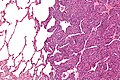
Monophasic synovial sarcoma with staghorn vessels - intermed. mag. (WC/Nephron)
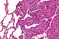
Monophasic synovial sarcoma with staghorn vessels - high mag. (WC/Nephron)

www:
- Synovial sarcoma (scielo.br).
- Synovial sarcoma - collection of images (humpath.com).
- Synovial sarcoma - several images (upmc.edu).
- Biphasic SS (radiographics.rsna.org).
- Monophasic SS (radiographics.rsna.org).
IHC
Features:[1]
- Vimentin +ve.
- EMA +ve.
- BCL2 +ve.
- CD99 +ve.
Others:
- Beta-catenin +ve ~30-70%.[6]
- Cyclin D1 ~50%.[6][7]
- TLE1 +ve nuclear staining; not specific for synovial sarcoma.[8][9]
Typically negative:[10]
- CD34.
- S100 ~30% +ve.
- SMA.
Notes:
- Synovial sarcoma & MPNST:
- Both +ve: PGP9.5 (UCHL1[11]), S100, NGFR, CD56, CD99, vimentin.
- Synovial +ve: EMA, keratin, BCL2, TLE1.
- MPNST +ve: nestin, CD34.
Trivia:
- PGP in PGP9.5 = protein gene product.[12]
EM
Features:[13]
- Biphasic tumour have biphasic ultrastructural features (unlike spindle cell carcinoma and epithelioid sarcoma).
- Epithelioid component is adenocarcinoma-like - they have:
- Intermediate filaments.
- Tonofilaments.
- Microvilli.
- Spindle cell component - mostly features less.
- Poorly formed desmosomes.
- No intermediate filaments, no myofilaments.
Molecular pathology
Associated translocation:
- t(X;18)(p11.2;q11.2).[14]
- SYT/SSX fusion gene.
Several SSX genes - cannot be differentiated with standard karyotyping:
- SSX1.
- SSX2 - better survival, rarely seen in biphasic tumours.[15]
- SSX4 - uncommon.
Notes:
- At HSC t(X,18) = synovial sarcoma.
See also
References
- ↑ 1.0 1.1 1.2 Humphrey, Peter A; Dehner, Louis P; Pfeifer, John D (2008). The Washington Manual of Surgical Pathology (1st ed.). Lippincott Williams & Wilkins. pp. 627. ISBN 978-0781765275.
- ↑ 2.0 2.1 2.2 Murphey, MD.; Gibson, MS.; Jennings, BT.; Crespo-Rodríguez, AM.; Fanburg-Smith, J.; Gajewski, DA.. "From the archives of the AFIP: Imaging of synovial sarcoma with radiologic-pathologic correlation.". Radiographics 26 (5): 1543-65. doi:10.1148/rg.265065084. PMID 16973781.
- ↑ URL: http://www.sarcomaimages.com/index.php?v=Synovial-Sarcoma. Accessed on: 2 April 2012.
- ↑ Schaal CH, Navarro FC, Moraes Neto FA (2004). "Primary renal sarcoma with morphologic and immunohistochemical aspects compatible with synovial sarcoma". Int Braz J Urol 30 (3): 210–3. PMID 15689250. http://www.brazjurol.com.br/may_june_2004/Schaal_ing_210_213.htm.
- ↑ Varela-Duran, J.; Enzinger, FM. (Jul 1982). "Calcifying synovial sarcoma.". Cancer 50 (2): 345-52. PMID 6282441.
- ↑ 6.0 6.1 Horvai AE, Kramer MJ, O'Donnell R (June 2006). "Beta-catenin nuclear expression correlates with cyclin D1 expression in primary and metastatic synovial sarcoma: a tissue microarray study". Arch. Pathol. Lab. Med. 130 (6): 792–8. PMID 16740029.
- ↑ Ng TL, Gown AM, Barry TS, et al. (January 2005). "Nuclear beta-catenin in mesenchymal tumors". Mod. Pathol. 18 (1): 68–74. doi:10.1038/modpathol.3800272. PMID 15375433.
- ↑ Kosemehmetoglu K, Vrana JA, Folpe AL (July 2009). "TLE1 expression is not specific for synovial sarcoma: a whole section study of 163 soft tissue and bone neoplasms". Mod. Pathol. 22 (7): 872–8. doi:10.1038/modpathol.2009.47. PMID 19363472. http://www.nature.com/modpathol/journal/v22/n7/full/modpathol200947a.html.
- ↑ Seo SW, Lee H, Lee HI, Kim HS (February 2011). "The role of TLE1 in synovial sarcoma". J Orthop Res. doi:10.1002/jor.21318. PMID 21319215.
- ↑ URL: http://path.upmc.edu/cases/case292/dx.html. Accessed on: 14 January 2012.
- ↑ Online 'Mendelian Inheritance in Man' (OMIM) 191342
- ↑ Doran, JF.; Jackson, P.; Kynoch, PA.; Thompson, RJ. (Jun 1983). "Isolation of PGP 9.5, a new human neurone-specific protein detected by high-resolution two-dimensional electrophoresis.". J Neurochem 40 (6): 1542-7. PMID 6343558.
- ↑ Fisher, C. (Dec 1998). "Synovial sarcoma.". Ann Diagn Pathol 2 (6): 401-21. PMID 9930576.
- ↑ URL: http://www.ncbi.nlm.nih.gov/omim/300813. Accessed on: 30 May 2010.
- ↑ Lefkowitch, Jay H. (2006). Anatomic Pathology Board Review (1st ed.). Saunders. pp. 625 Q6. ISBN 978-1416025887.