Subependymoma
Jump to navigation
Jump to search
The printable version is no longer supported and may have rendering errors. Please update your browser bookmarks and please use the default browser print function instead.
| Subependymoma | |
|---|---|
| Diagnosis in short | |
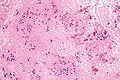
 Subependymoma. H&E stain. | |
|
| |
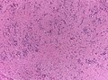
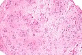
| LM | microcysts with bluish material (give a spongy appearance at low magnification), clustering of nuclei cluster (described as "bundles of flowers"), bland nuclei |
| Site | brain - see neuropathology tumours |
|
| |
| Symptoms | +/-headache |
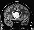
| Radiology | classically fourth ventricle |
| Prognosis | WHO grade I |
| Clin. DDx | other brain tumours - ependymoma, CNS lymphoma |
| Treatment | surgical excision |
Subependymoma is neuropathology tumour classically found in the fourth ventricle.
General
- Good prognosis - WHO Grade I (ICD-O 9383/1).
- Low-grade glial tumour of the middle-aged and elderly.[1]
- Formerly called subependymal (glomerate) astrocytoma
Clinical:[1]
- Slow growing.
- +/-Headaches.
- CSF obstructions.
- Tx: surgery.
Gross/radiology
- Classic location: fourth ventricle (50-60%).[2]
- Lateral ventricles (30-40% of all cases), rarely IIIrd ventricle, septum pellucideum and spinal cord
- Well-demarcated margin.
- Usu. completely within the ventricle; does not extend into brain (like ependymomas).
- May be hemorrhagic. [3]
- Usually less than 2cm in diameter.
- Incidentally found at autopsies.
Images
Subependymoma (AFIP)
Macroscopy (AFIP)


Microscopic
Features:[4]
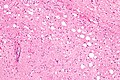
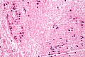
- Microcysts with bluish material - give a spongy appearance at low magnification.
- Isomorphic nuclei cluster embedded into fibrillary matrix.
- Described as "bundles of flowers".
- Calcifications possible
- Combined subependymomas/classical cellular ependymomas (then grade II)
Negatives.
- No nuclear pleomorphism, no prominent nucleoli, no mitoses.
- Do not invade into brain.[1]
Images
www: Subependymoma (ouhsc.edu).[4]
Subependymoma - low mag. (WC/jensflorian)
Subependyoma - intermed. mag. (WC)
Subependymoma - high mag. (WC)
Subependymoma - high mag. (WC/Nephron)
Subependymoma - very high mag. (WC)
Subependymoma (AFIP)


DDx: Ependymoma
IHC
- GFAP+ve
- MIB-1 very low (1%)
- ATRX: no loss
- IDH1(R132H)-ve
Molecular
- Posterior fossa and spine: chr.6 alterations.
- Supratentorial: No consistent abberations.
See also
References
- ↑ 1.0 1.1 1.2 Castro-Castro, J.; Castro-Bouzas, D.; Prieto-Casal, PL.; Carcacia-Hermilla, ID.; Riu-Lloveras, M.; Castro-Gómez, JE. (Mar 2013). "[Subependymoma of the lateral ventricle. A case report].". Rev Neurol 56 (6): 332-6. PMID 23483468.
- ↑ Hoeffel, C.; Boukobza, M.; Polivka, M.; Lot, G.; Guichard, JP.; Lafitte, F.; Reizine, D.; Merland, JJ.. "MR manifestations of subependymomas.". AJNR Am J Neuroradiol 16 (10): 2121-9. PMID 8585504. http://www.ajnr.org/cgi/reprint/16/10/2121.
- ↑ Landriel, F.; Besada, C.; Migliaro, M.; Christiansen, S.; Goldschmidt, E.; Yampolsky, C.; Ajler, P. (2013). "Atypical hemorrhagic presentation of a fourth ventricle subependymoma: case report.". Neurol Med Chir (Tokyo) 53 (11): 828-31. PMID 24140775.
- ↑ 4.0 4.1 URL: http://moon.ouhsc.edu/kfung/jty1/Com05/Com501-2-Diss.htm. Accessed on: 2 June 2011.