Difference between revisions of "Subependymoma"
Jump to navigation
Jump to search
Jensflorian (talk | contribs) (update + pictures from WC) |
|||
| Line 33: | Line 33: | ||
==General== | ==General== | ||
*Good prognosis - WHO Grade I. | *Good prognosis - WHO Grade I (ICD-O 9383/1). | ||
*Low-grade glial tumour.<ref name=pmid23483468/> | *Low-grade glial tumour of the middle-aged and elderly.<ref name=pmid23483468/> | ||
*Formerly called ''subependymal (glomerate) astrocytoma'' | |||
Clinical:<ref name=pmid23483468>{{Cite journal | last1 = Castro-Castro | first1 = J. | last2 = Castro-Bouzas | first2 = D. | last3 = Prieto-Casal | first3 = PL. | last4 = Carcacia-Hermilla | first4 = ID. | last5 = Riu-Lloveras | first5 = M. | last6 = Castro-Gómez | first6 = JE. | title = [Subependymoma of the lateral ventricle. A case report]. | journal = Rev Neurol | volume = 56 | issue = 6 | pages = 332-6 | month = Mar | year = 2013 | doi = | PMID = 23483468 }} | Clinical:<ref name=pmid23483468>{{Cite journal | last1 = Castro-Castro | first1 = J. | last2 = Castro-Bouzas | first2 = D. | last3 = Prieto-Casal | first3 = PL. | last4 = Carcacia-Hermilla | first4 = ID. | last5 = Riu-Lloveras | first5 = M. | last6 = Castro-Gómez | first6 = JE. | title = [Subependymoma of the lateral ventricle. A case report]. | journal = Rev Neurol | volume = 56 | issue = 6 | pages = 332-6 | month = Mar | year = 2013 | doi = | PMID = 23483468 }} | ||
| Line 40: | Line 41: | ||
*Slow growing. | *Slow growing. | ||
*+/-Headaches. | *+/-Headaches. | ||
*CSF obstructions. | |||
*Tx: surgery. | *Tx: surgery. | ||
==Gross/radiology== | ==Gross/radiology== | ||
*Classic location: fourth ventricle.<ref>{{Cite journal | last1 = Hoeffel | first1 = C. | last2 = Boukobza | first2 = M. | last3 = Polivka | first3 = M. | last4 = Lot | first4 = G. | last5 = Guichard | first5 = JP. | last6 = Lafitte | first6 = F. | last7 = Reizine | first7 = D. | last8 = Merland | first8 = JJ. | title = MR manifestations of subependymomas. | journal = AJNR Am J Neuroradiol | volume = 16 | issue = 10 | pages = 2121-9 | month = | year = | doi = | PMID = 8585504 |url=http://www.ajnr.org/cgi/reprint/16/10/2121}}</ref> | *Classic location: fourth ventricle (50-60%).<ref>{{Cite journal | last1 = Hoeffel | first1 = C. | last2 = Boukobza | first2 = M. | last3 = Polivka | first3 = M. | last4 = Lot | first4 = G. | last5 = Guichard | first5 = JP. | last6 = Lafitte | first6 = F. | last7 = Reizine | first7 = D. | last8 = Merland | first8 = JJ. | title = MR manifestations of subependymomas. | journal = AJNR Am J Neuroradiol | volume = 16 | issue = 10 | pages = 2121-9 | month = | year = | doi = | PMID = 8585504 |url=http://www.ajnr.org/cgi/reprint/16/10/2121}}</ref> | ||
*Lateral ventricles (30-40% of all cases), rarely IIIrd ventricle, septum pellucideum and spinal cord | |||
*Well-demarcated margin. | *Well-demarcated margin. | ||
*Usu. completely within the ventricle; does not extend into brain (like [[ependymoma]]s). | *Usu. completely within the ventricle; does not extend into brain (like [[ependymoma]]s). | ||
*May be hemorrhagic. <ref>{{Cite journal | last1 = Landriel | first1 = F. | last2 = Besada | first2 = C. | last3 = Migliaro | first3 = M. | last4 = Christiansen | first4 = S. | last5 = Goldschmidt | first5 = E. | last6 = Yampolsky | first6 = C. | last7 = Ajler | first7 = P. | title = Atypical hemorrhagic presentation of a fourth ventricle subependymoma: case report. | journal = Neurol Med Chir (Tokyo) | volume = 53 | issue = 11 | pages = 828-31 | month = | year = 2013 | doi = | PMID = 24140775 }}</ref> | |||
*Usually less than 2cm in diameter. | |||
*Incidentally found at autopsies. | |||
===Images=== | |||
<gallery> | |||
File:AFIP405740R-SUBEPENDYMOMA.jpg | Subependymoma (AFIP) | |||
File:Subependymoma.jpg | Macroscopy (AFIP) | |||
</gallery> | |||
==Microscopic== | ==Microscopic== | ||
Features:<ref name=ouhsc>URL: [http://moon.ouhsc.edu/kfung/jty1/Com05/Com501-2-Diss.htm http://moon.ouhsc.edu/kfung/jty1/Com05/Com501-2-Diss.htm]. Accessed on: 2 June 2011.</ref> | Features:<ref name=ouhsc>URL: [http://moon.ouhsc.edu/kfung/jty1/Com05/Com501-2-Diss.htm http://moon.ouhsc.edu/kfung/jty1/Com05/Com501-2-Diss.htm]. Accessed on: 2 June 2011.</ref> | ||
*Microcysts with bluish material - give a spongy appearance at low magnification. | *Microcysts with bluish material - give a spongy appearance at low magnification. | ||
* | *Isomorphic nuclei cluster embedded into fibrillary matrix. | ||
**Described as "bundles of flowers". | **Described as "bundles of flowers". | ||
*Calcifications possible | |||
*Combined subependymomas/classical cellular ependymomas (then grade II) | |||
Negatives. | Negatives. | ||
| Line 58: | Line 72: | ||
===Images=== | ===Images=== | ||
www: | www: [http://moon.ouhsc.edu/kfung/jty1/Com05/Com05Image/Com501-2-04.gif Subependymoma (ouhsc.edu)].<ref name=ouhsc>URL: [http://moon.ouhsc.edu/kfung/jty1/Com05/Com501-2-Diss.htm http://moon.ouhsc.edu/kfung/jty1/Com05/Com501-2-Diss.htm]. Accessed on: 2 June 2011.</ref> | ||
<gallery> | <gallery> | ||
File:Subependymoma HE x40.jpg | Subependymoma - low mag. (WC/jensflorian) | |||
Image:Subependymoma_-_intermed_mag.jpg | Subependyoma - intermed. mag. (WC) | Image:Subependymoma_-_intermed_mag.jpg | Subependyoma - intermed. mag. (WC) | ||
Image:Subependymoma_-_high_mag.jpg | Subependymoma - high mag. (WC) | Image:Subependymoma_-_high_mag.jpg | Subependymoma - high mag. (WC) | ||
File:Subependymoma 2 - high mag.jpg | Subependymoma - high mag. (WC/Nephron) | |||
Image:Subependymoma_-_very_high_mag.jpg | Subependymoma - very high mag. (WC) | Image:Subependymoma_-_very_high_mag.jpg | Subependymoma - very high mag. (WC) | ||
File:AFIP405743M-SUBEPENDYMOMA.jpg | Subependymoma (AFIP) | |||
</gallery> | </gallery> | ||
DDx: | |||
[[Ependymoma]] | |||
==IHC== | |||
*GFAP+ve | |||
*MIB-1 very low (1%) | |||
*ATRX: no loss | |||
*IDH1(R132H)-ve | |||
==Molecular== | |||
*No consistent abberations | |||
==See also== | ==See also== | ||
Revision as of 15:02, 15 April 2015
| Subependymoma | |
|---|---|
| Diagnosis in short | |
 Subependymoma. H&E stain. | |
|
| |
| LM | microcysts with bluish material (give a spongy appearance at low magnification), clustering of nuclei cluster (described as "bundles of flowers"), bland nuclei |
| Site | brain - see neuropathology tumours |
|
| |
| Symptoms | +/-headache |
| Radiology | classically fourth ventricle |
| Prognosis | WHO grade I |
| Clin. DDx | other brain tumours - ependymoma, CNS lymphoma |
| Treatment | surgical excision |
Subependymoma is neuropathology tumour classically found in the fourth ventricle.
General
- Good prognosis - WHO Grade I (ICD-O 9383/1).
- Low-grade glial tumour of the middle-aged and elderly.[1]
- Formerly called subependymal (glomerate) astrocytoma
Clinical:[1]
- Slow growing.
- +/-Headaches.
- CSF obstructions.
- Tx: surgery.
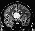
Gross/radiology
- Classic location: fourth ventricle (50-60%).[2]
- Lateral ventricles (30-40% of all cases), rarely IIIrd ventricle, septum pellucideum and spinal cord
- Well-demarcated margin.
- Usu. completely within the ventricle; does not extend into brain (like ependymomas).
- May be hemorrhagic. [3]
- Usually less than 2cm in diameter.
- Incidentally found at autopsies.
Images
Subependymoma (AFIP)
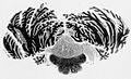
Macroscopy (AFIP)


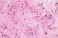
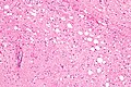
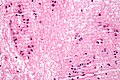
Microscopic
Features:[4]
- Microcysts with bluish material - give a spongy appearance at low magnification.
- Isomorphic nuclei cluster embedded into fibrillary matrix.
- Described as "bundles of flowers".
- Calcifications possible
- Combined subependymomas/classical cellular ependymomas (then grade II)
Negatives.
- No nuclear pleomorphism, no prominent nucleoli, no mitoses.
- Do not invade into brain.[1]
Images
www: Subependymoma (ouhsc.edu).[4]
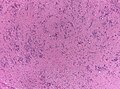
Subependymoma - low mag. (WC/jensflorian)
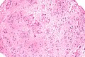
Subependyoma - intermed. mag. (WC)
Subependymoma - high mag. (WC)
Subependymoma - high mag. (WC/Nephron)
Subependymoma - very high mag. (WC)
Subependymoma (AFIP)


DDx: Ependymoma
IHC
- GFAP+ve
- MIB-1 very low (1%)
- ATRX: no loss
- IDH1(R132H)-ve
Molecular
- No consistent abberations
See also
References
- ↑ 1.0 1.1 1.2 Castro-Castro, J.; Castro-Bouzas, D.; Prieto-Casal, PL.; Carcacia-Hermilla, ID.; Riu-Lloveras, M.; Castro-Gómez, JE. (Mar 2013). "[Subependymoma of the lateral ventricle. A case report].". Rev Neurol 56 (6): 332-6. PMID 23483468.
- ↑ Hoeffel, C.; Boukobza, M.; Polivka, M.; Lot, G.; Guichard, JP.; Lafitte, F.; Reizine, D.; Merland, JJ.. "MR manifestations of subependymomas.". AJNR Am J Neuroradiol 16 (10): 2121-9. PMID 8585504. http://www.ajnr.org/cgi/reprint/16/10/2121.
- ↑ Landriel, F.; Besada, C.; Migliaro, M.; Christiansen, S.; Goldschmidt, E.; Yampolsky, C.; Ajler, P. (2013). "Atypical hemorrhagic presentation of a fourth ventricle subependymoma: case report.". Neurol Med Chir (Tokyo) 53 (11): 828-31. PMID 24140775.
- ↑ 4.0 4.1 URL: http://moon.ouhsc.edu/kfung/jty1/Com05/Com501-2-Diss.htm. Accessed on: 2 June 2011.