Steatocystoma
Jump to navigation
Jump to search
The printable version is no longer supported and may have rendering errors. Please update your browser bookmarks and please use the default browser print function instead.
| Steatocystoma | |
|---|---|
| Diagnosis in short | |
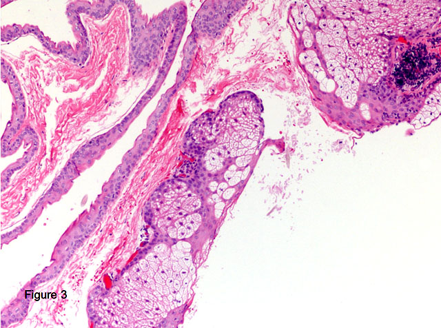
 Steatocystoma. H&E stain. | |
|
| |
| LM | cyst lined by squamous epithelium with a corrugated eosinophilic lining, no granular cell layer |
| Site | skin - see dermal cysts |
|
| |
| Syndromes | steatocystoma multiplex |
|
| |
| Prevalence | rare |
| Prognosis | benign |
Steatocystoma is a rare benign dermal cyst.
General
- Benign.
- Rare.[1]
- Typically adults.
- Usually on the trunk.
- May be genetic; known as steatocystoma multiplex.[2]
- Classically autosomal dominant.[3]
Microscopic
Features:[4]
- Cyst lined by squamous epithelium with:
- Corrugated eosinophilic lining - key feature.
- Similar appearance to compact keratin (hyperkeratosis).
- Described as a hyaline cuticle.[5]
- No granular cell layer.
- Corrugated eosinophilic lining - key feature.
Images
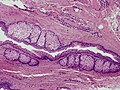
Steatocystoma. (WC)
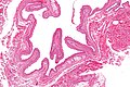
Steatocystoma - low mag. (WC/Nephron)
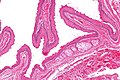
Steatocystoma - intermed. mag. (WC/Nephron)
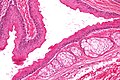
Steatocystoma - high mag. (WC/Nephron)


www:
See also
References
- ↑ Gordon Spratt, EA.; Kaplan, J.; Patel, RR.; Kamino, H.; Ramachandran, SM. (Dec 2013). "Steatocystoma.". Dermatol Online J 19 (12): 20721. PMID 24365012.
- ↑ Online 'Mendelian Inheritance in Man' (OMIM) 184500
- ↑ URL: http://path.upmc.edu/cases/case674/dx.html. Accessed on: 29 January 2012.
- ↑ Busam, Klaus J. (2009). Dermatopathology: A Volume in the Foundations in Diagnostic Pathology Series (1st ed.). Saunders. pp. 312. ISBN 978-0443066542.
- ↑ URL: http://path.upmc.edu/cases/case674/dx.html. Accessed on: 29 January 2012.
- ↑ URL: http://path.upmc.edu/cases/case674.html. Accessed on: 29 January 2012.