Spindle cell
Jump to navigation
Jump to search
The printable version is no longer supported and may have rendering errors. Please update your browser bookmarks and please use the default browser print function instead.


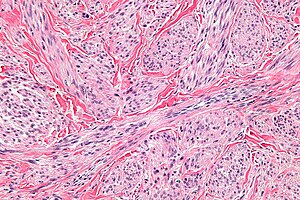
Spindle cells in a leiomyosarcoma. (WC)
Spindle cell is a histomorphologic descriptor used in pathology.
A list of spindle cell lesions is found the in the spindle cell lesions article.
Definition
It refers to a cell that is tapered at both ends.[1]
Notes:
- A taper gradually decreases toward one end [of the cross-section or width].[2]
- Image: Taperred thread (qcfocus.com).
- Spindle cells can have "pointy" ends (typical for nerves) or "rounded" ends (typical for muscle), i.e. be ellipitcal or vesica piscis.
Subtyping spindle cells by H&E
Spindle cells can often be subtyped based on H&E:[3]
- Fibroblast = blue.
- Smooth muscle = deep pink.
- Myofibroblast = purple.
Images
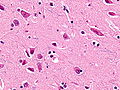
Spindle neurons. (WC)
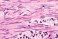
Benign smooth muscle cells of the urinary bladder. (WC)
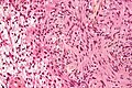
Spindle cells of a schwannoma. (WC)


Shapes

A spindle. (WC)

Vesica piscis. (WC)
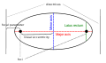
Ellipse. (WC)


See also
References
- ↑ URL: http://www.medterms.com/script/main/art.asp?articlekey=25657. Accessed on: 2 February 2011.
- ↑ URL: http://dictionary.reference.com/browse/taper. Accessed on: 3 February 2011.
- ↑ Chan, JK. (Feb 2014). "The wonderful colors of the hematoxylin-eosin stain in diagnostic surgical pathology.". Int J Surg Pathol 22 (1): 12-32. doi:10.1177/1066896913517939. PMID 24406626.