Difference between revisions of "Solitary fibrous tumour"
m (→Microscopic) |
m (→Images) |
||
| Line 75: | Line 75: | ||
[[File:3 18039322534203 sl 4.png|malignant solitary fibrous tumor]] | [[File:3 18039322534203 sl 4.png|malignant solitary fibrous tumor]] | ||
Malignant solitary fibrous tumor (low to intermediate grade tumor). 15 cm mass in upper arm of 50 year old man. A. Hypocellular (green arrow) and hypercellular (cyan arrow) areas alternate. B. Vascular spaces suggest antlers (staghorn). Note subcapsular hemorrhage. C. Stromal hyalinization. D. Nuclei, lacking organization, show varied size and shape, with some cells being round to ovoid. The arrow points to myxoid focus in stroma. When a majority of the following are present, a low to intermediate grade malignant solitary fibrous tumor is present: 1) size > 5 cm (this case), 2) pleomorphism with round cells/epithelioid cells (this case), 3) subcapsular hemorrhage (this case), 4) ≥4 mitoses per 10 high power fields, 5) necrosis with perivascular tumor sparing. STAT 6 positivity is very helpful in separating this tumor from its mimics. Am J Surg Pathol | Malignant solitary fibrous tumor (low to intermediate grade tumor). 15 cm mass in upper arm of 50 year old man. A. Hypocellular (green arrow) and hypercellular (cyan arrow) areas alternate. B. Vascular spaces suggest antlers (staghorn). Note subcapsular hemorrhage. C. Stromal hyalinization. D. Nuclei, lacking organization, show varied size and shape, with some cells being round to ovoid. The arrow points to myxoid focus in stroma. When a majority of the following are present, a low to intermediate grade malignant solitary fibrous tumor is present: 1) size > 5 cm (this case), 2) pleomorphism with round cells/epithelioid cells (this case), 3) subcapsular hemorrhage (this case), 4) ≥4 mitoses per 10 high power fields, 5) necrosis with perivascular tumor sparing. STAT 6 positivity is very helpful in separating this tumor from its mimics. <ref name=pmid24625420>{{cite journal |author=Yoshida A1, Tsuta K, Ohno M, Yoshida M, Narita Y, Kawai A, Asamura H, Kushima R |title=STAT6 immunohistochemistry is helpful in the diagnosis of solitary fibrous tumors |journal=Am J Surg Pathol |volume=38 |issue=4 |pages=552-559 |year=2014 |month=April |pmid=24625420 |doi=10.1097/PAS.0000000000000137}}</ref> | ||
==IHC== | ==IHC== | ||
Revision as of 20:41, 14 December 2016
| Solitary fibrous tumour | |
|---|---|
| Diagnosis in short | |
 Solitary fibrous tumour. H&E stain. | |
|
| |
| LM | spindle cells in a patternless pattern, hemangiopericytoma-like areas (staghorn vessels), keloid-like collagen bundles, +/-well-circumscribed (common) |
| Subtypes | benign (common), malignant (uncommon) |
| IHC | CD34 ~90% +ve, CD99 ~70% +ve, BCL2 ~50% +ve |
| Site | soft tissue - fibroblastic/myofibroblastic tumours, pleura |
|
| |
| Syndromes | Doege-Potter syndrome |
|
| |
| Prognosis | usu. good |
Solitary fibrous tumour, abbreviated SFT, is a type of soft tissue tumour that fits in the fibroblastic/myofibroblastic tumours. It is usually benign.
SFT of the pleura is dealt with in a separate article solitary fibrous tumour of the pleura.
General
- Grouped with hemangiopericytoma in the WHO classification - as it is thought to be the same tumour because both share the same molecular alteration.[1][2]
- May be benign or malignant; more commonly benign.[3][4]
- May be associated with hypoglycemia.
- Known as Doege-Potter syndrome.[5]
- Leptomeningeal SFTs/hemangiopericytomas are classified as follows:
- WHO grade I: classical SFT
- WHO grade II: classical hemangiopericytoma
- WHO grade III: anaplastic hemangiopericytoma / malignant SFT
Gross
- Soft tissue mass.
Microscopic
Features - benign:
- Spindle cells in a patternless pattern.
- Occasionally epithelioid cells - rare.[6]
- Hemangiopericytoma-like area (staghorn vessels).
- Keloid-like collagen bundles - key feature.
- +/-Well-circumscribed (common).
Criteria for malignancy:[1]
- Necrosis.
- Mitoses >4/10 HPF -- definition suffers from HPFitis.
- Increased cellularity.
- Marked nuclear atypia.
- Infiltrative margin.
Images
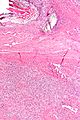
Benign SFT - low mag. (WC)
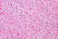
Benign SFT - intermed. mag. (WC)
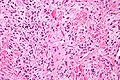
Benign SFT - high mag. (WC)


www:


Malignant solitary fibrous tumor (low to intermediate grade tumor). 15 cm mass in upper arm of 50 year old man. A. Hypocellular (green arrow) and hypercellular (cyan arrow) areas alternate. B. Vascular spaces suggest antlers (staghorn). Note subcapsular hemorrhage. C. Stromal hyalinization. D. Nuclei, lacking organization, show varied size and shape, with some cells being round to ovoid. The arrow points to myxoid focus in stroma. When a majority of the following are present, a low to intermediate grade malignant solitary fibrous tumor is present: 1) size > 5 cm (this case), 2) pleomorphism with round cells/epithelioid cells (this case), 3) subcapsular hemorrhage (this case), 4) ≥4 mitoses per 10 high power fields, 5) necrosis with perivascular tumor sparing. STAT 6 positivity is very helpful in separating this tumor from its mimics. [7]
IHC
- CD34 ~90% +ve.
- CD99 ~70% +ve.
- BCL2 ~50% +ve.
- Stat6 nuclear +ve.[8]
Molecular
DDx
- Meningioma
- Cellular angiofibroma
- Myofibroblastoma
- Benign fibrous histiocytoma
- Dermatofibrosarcoma protruberans
- Fibromyxoid sarcoma
See also
References
- ↑ 1.0 1.1 Humphrey, Peter A; Dehner, Louis P; Pfeifer, John D (2008). The Washington Manual of Surgical Pathology (1st ed.). Lippincott Williams & Wilkins. pp. 609. ISBN 978-0781765275.
- ↑ Schweizer, L.; Koelsche, C.; Sahm, F.; Piro, RM.; Capper, D.; Reuss, DE.; Pusch, S.; Habel, A. et al. (May 2013). "Meningeal hemangiopericytoma and solitary fibrous tumors carry the NAB2-STAT6 fusion and can be diagnosed by nuclear expression of STAT6 protein.". Acta Neuropathol 125 (5): 651-8. doi:10.1007/s00401-013-1117-6. PMID 23575898.
- ↑ URL: http://www.pathconsultddx.com/pathCon/diagnosis?pii=S1559-8675%2806%2970528-9. Accessed on: 25 June 2010.
- ↑ URL: http://wjso.com/content/6/1/86. Accessed on: 25 June 2010.
- ↑ Roy, TM.; Burns, MV.; Overly, DJ.; Curd, BT. (Nov 1992). "Solitary fibrous tumor of the pleura with hypoglycemia: the Doege-Potter syndrome.". J Ky Med Assoc 90 (11): 557-60. PMID 1474302.
- ↑ Martorell, M.; Pérez-Vallés, A.; Gozalbo, F.; Garcia-Garcia, JA.; Gutierrez, J.; Gaona, J. (2007). "Solitary fibrous tumor of the thigh with epithelioid features: a case report.". Diagn Pathol 2: 19. doi:10.1186/1746-1596-2-19. PMID 17577399.
- ↑ Yoshida A1, Tsuta K, Ohno M, Yoshida M, Narita Y, Kawai A, Asamura H, Kushima R (April 2014). "STAT6 immunohistochemistry is helpful in the diagnosis of solitary fibrous tumors". Am J Surg Pathol 38 (4): 552-559. doi:10.1097/PAS.0000000000000137. PMID 24625420.
- ↑ Cheah, AL.; Billings, SD.; Goldblum, JR.; Carver, P.; Tanas, MZ.; Rubin, BP. (Aug 2014). "STAT6 rabbit monoclonal antibody is a robust diagnostic tool for the distinction of solitary fibrous tumour from its mimics.". Pathology 46 (5): 389-95. doi:10.1097/PAT.0000000000000122. PMID 24977739.
- ↑ Mohajeri, A.; Tayebwa, J.; Collin, A.; Nilsson, J.; Magnusson, L.; von Steyern, FV.; Brosjö, O.; Domanski, HA. et al. (Oct 2013). "Comprehensive genetic analysis identifies a pathognomonic NAB2/STAT6 fusion gene, nonrandom secondary genomic imbalances, and a characteristic gene expression profile in solitary fibrous tumor.". Genes Chromosomes Cancer 52 (10): 873-86. doi:10.1002/gcc.22083. PMID 23761323.
- ↑ Robinson, DR.; Wu, YM.; Kalyana-Sundaram, S.; Cao, X.; Lonigro, RJ.; Sung, YS.; Chen, CL.; Zhang, L. et al. (Feb 2013). "Identification of recurrent NAB2-STAT6 gene fusions in solitary fibrous tumor by integrative sequencing.". Nat Genet 45 (2): 180-5. doi:10.1038/ng.2509. PMID 23313952.