Difference between revisions of "Serous cystadenoma of the pancreas"
Jump to navigation
Jump to search
(redirect) |
(split -out) |
||
| Line 1: | Line 1: | ||
'''Serous cystadenoma of the pancreas''' is a benign cystic lesion of the pancreas. It may be seen in [[von Hippel-Lindau disease]]. | |||
It is also known as '''serous microcystic adenoma''',<ref name=Ref_Sternberg4_1630>{{Ref Sternberg4|1630}}</ref> and '''pancreatic serous cystadenoma'''. | |||
==General== | |||
*1-2% of all exocrine pancreatic tumours. | |||
*Female > male. | |||
*Mean age 66 years. | |||
*Truly benign with no malignant potenial. | |||
*May be part of [[von Hippel-Lindau syndrome]]. | |||
Management: | |||
*Observe or resect. | |||
==Gross== | |||
Features: | |||
*Classically has a characteristic central scar.<ref name=pmid15888617>{{cite journal |author=Kim YH, Saini S, Sahani D, Hahn PF, Mueller PR, Auh YH |title=Imaging diagnosis of cystic pancreatic lesions: pseudocyst versus nonpseudocyst |journal=Radiographics |volume=25 |issue=3 |pages=671–85 |year=2005 |pmid=15888617 |doi=10.1148/rg.253045104 |url=http://radiographics.rsna.org/content/25/3/671.abstract}}</ref> | |||
*Bosulated surface. | |||
*Lobulated. | |||
*No (macroscopic) cysts apparent on gross. | |||
*Location: 50-70% occur in the body and tail. | |||
*Size: average size 11 cm. | |||
Radiologic appearance: | |||
*Honey comb-like appearance. | |||
*Well demarcated border - may be described as a "coin lesion". | |||
Image: | |||
*[http://www.joplink.net/prev/200905/25_fig06.jpg Serous microcystic adenoma (joplink.net)].<ref name=pmid19454830>{{Cite journal | last1 = Vernadakis | first1 = S. | last2 = Kaiser | first2 = GM. | last3 = Christodoulou | first3 = E. | last4 = Mathe | first4 = Z. | last5 = Troullinakis | first5 = M. | last6 = Bankfalvi | first6 = A. | last7 = Paul | first7 = A. | title = Enormous serous microcystic adenoma of the pancreas. | journal = JOP | volume = 10 | issue = 3 | pages = 332-4 | month = | year = 2009 | doi = | PMID = 19454830 |URL = http://www.joplink.net/prev/200905/25.html }}</ref> | |||
==Microscopic== | |||
Features: | |||
*Cystic spaces lined by cuboidal cells. | |||
**Glycogen rich. | |||
**Cilia. (???) | |||
DDx: | |||
*[[Renal cell carcinoma]]. | |||
*[[Lymphangioma]]. | |||
*[[Hemangioma]]. | |||
*Oligocystic mucinous cystic tumors and pseudocysts. | |||
**Have mucin; PAS-D could be used to demonstrate its presence. | |||
Notes: | |||
*Serous adenoma may coexist with aggressive tumours. | |||
===Images=== | |||
<gallery> | |||
Image:Pancreatic_serous_cystadenoma_-_low_mag.jpg | PSC - low mag. (WC) | |||
Image:Pancreatic_serous_cystadenoma_-_intermed_mag.jpg | PSC - intermed. mag. (WC) | |||
Image:Pancreatic_serous_cystadenoma_-_high_mag.jpg | PSC - high mag. (WC) | |||
Image:Pancreatic_serous_cystadenoma_-_very_high_mag.jpg | PSC - very high mag. (WC) | |||
</gallery> | |||
==Stains== | |||
*PAS +ve. | |||
*PASD -ve. | |||
==See also== | |||
*[[Pancreas]]. | |||
*[[Serous cystadenoma]]. | |||
==References== | |||
{{Reflist|2}} | |||
[[Category:Pancreas]] | |||
[[Category:Diagnosis]] | [[Category:Diagnosis]] | ||
Revision as of 01:26, 18 January 2014
Serous cystadenoma of the pancreas is a benign cystic lesion of the pancreas. It may be seen in von Hippel-Lindau disease.
It is also known as serous microcystic adenoma,[1] and pancreatic serous cystadenoma.
General
- 1-2% of all exocrine pancreatic tumours.
- Female > male.
- Mean age 66 years.
- Truly benign with no malignant potenial.
- May be part of von Hippel-Lindau syndrome.
Management:
- Observe or resect.
Gross
Features:
- Classically has a characteristic central scar.[2]
- Bosulated surface.
- Lobulated.
- No (macroscopic) cysts apparent on gross.
- Location: 50-70% occur in the body and tail.
- Size: average size 11 cm.
Radiologic appearance:
- Honey comb-like appearance.
- Well demarcated border - may be described as a "coin lesion".
Image:
Microscopic
Features:
- Cystic spaces lined by cuboidal cells.
- Glycogen rich.
- Cilia. (???)
DDx:
- Renal cell carcinoma.
- Lymphangioma.
- Hemangioma.
- Oligocystic mucinous cystic tumors and pseudocysts.
- Have mucin; PAS-D could be used to demonstrate its presence.
Notes:
- Serous adenoma may coexist with aggressive tumours.
Images
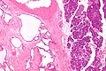
PSC - low mag. (WC)
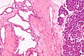
PSC - intermed. mag. (WC)
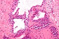
PSC - high mag. (WC)
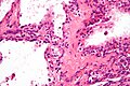
PSC - very high mag. (WC)


Stains
- PAS +ve.
- PASD -ve.
See also
References
- ↑ Mills, Stacey E; Carter, Darryl; Greenson, Joel K; Oberman, Harold A; Reuter, Victor E (2004). Sternberg's Diagnostic Surgical Pathology (4th ed.). Lippincott Williams & Wilkins. pp. 1630. ISBN 978-0781740517.
- ↑ Kim YH, Saini S, Sahani D, Hahn PF, Mueller PR, Auh YH (2005). "Imaging diagnosis of cystic pancreatic lesions: pseudocyst versus nonpseudocyst". Radiographics 25 (3): 671–85. doi:10.1148/rg.253045104. PMID 15888617. http://radiographics.rsna.org/content/25/3/671.abstract.
- ↑ Vernadakis, S.; Kaiser, GM.; Christodoulou, E.; Mathe, Z.; Troullinakis, M.; Bankfalvi, A.; Paul, A. (2009). "Enormous serous microcystic adenoma of the pancreas.". JOP 10 (3): 332-4. PMID 19454830.